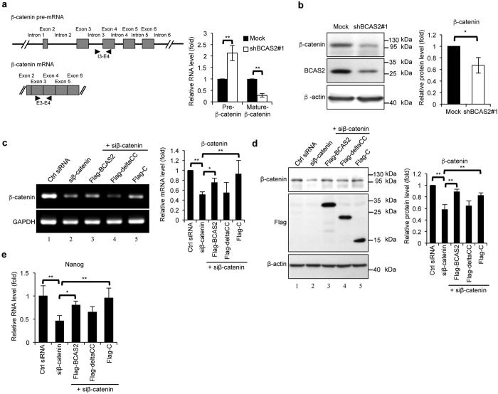
Figure 1. BCAS2 regulates β-catenin gene expression.
(a) BCAS2 knockdown in MCF7 cells increases β-catenin pre-mRNA and reduces β-catenin mRNA levels. Left: schematic representation of the design of primers to detect the intron-containing precursor mRNA (upper) and mRNA of β-catenin (lower). The locations of primers, exons and introns are denoted with arrowheads, boxes and lines, respectively. Right: quantitative RT-PCR analysis (n = 3). (b) Decreased β-catenin protein in BCAS2-knockdown MCF7 cells. Left, a representative western blot. Right, quantification of the left panel (n = 3). (c,d) Full-length BCAS2 and C-terminal of BCAS2 increased the β-catenin RNA (c) and protein (d) expression in siβ-catenin-treated N2A cells, but deltaCC did not affect the β-catenin expression. The full-length BCAS2, C-fragment, and deltaCC mutant plasmids were transfected into the β-catenin-silenced N2A cells separately. Forty-eight hours after transfection, the RNA and protein were extracted for RT-PCR and western analysis, respectively. Left: representative result. Right: quantitation of left panel (n = 3). (e) qRT-PCR showing nanog expression (n = 3). Panels a, b: The P value was analyzed by a two-tailed Student’s t-test. Panels c, d, and e: one-way ANOVA. Results are shown as the mean ± S.D. Uncropped blots are presented in Supplementary Fig. S5.

