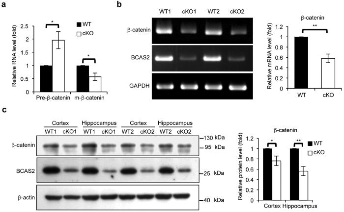
Figure 7. Reduced β-catenin gene expression in the BCAS2 cKO hippocampus.
(a) RNA was extracted from the hippocampus of WT and BCAS2 cKO mice. Quantitative RT-PCR analysis showed that loss of BCAS2 caused increased β-catenin pre-mRNA and reduced β-catenin mRNA levels, as evaluated by three independent experiments, indicating the impaired splicing efficiency of β-catenin pre-mRNA (n = 3). (b) RT-PCR analysis of the β-catenin expression level. Left, representative result. Right, quantification of the left panel (n = 3). (c) Western blot analysis of the protein levels of BCAS2 and β-catenin in the cortex and hippocampus of each genotype. Left, representative western blot. Right, quantification of the left panel (n = 3). Results are shown as the mean ± S.D. and analyzed by a two-tailed Student’s test. Uncropped blots are presented in Supplementary Fig. S5.

