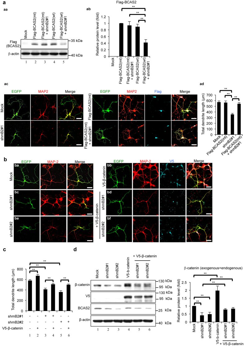
Figure 8. Overexpression of β-catenin can rescue the impaired dendritic growth of the BCAS2 knockdown in a primary neuron culture.
(a) Identification of shBCAS2 to target mouse BCAS2 gene. (aa) Western blot analysis. shmB2#1 could not reduced the exogenous BCAS2 expression while co-transfected with BCAS2 silent mutant (Flag-BCAS2(mt)). (ab) Quantification of three independent experiments analyzed by one-way ANOVA. (ac) BCAS2 silent mutant can rescue dendrite length in shRNA treated primary neuron cells. The primary neurons isolated from the forebrains of E18.5 embryos were seeded and then transfected with the following: the pLL3.7 (Mock), shRNAs targeting BCAS2 (shmB2#1) or Flag-tagged BCAS2(mt) alone, or shBCAS2 plasmid with Flag-tagged BCAS2(mt). IFA was performed 5 days after the plasmid transfection using anti-MAP-2 and anti-Flag antibodies. Dendritic length was measured from 30 merged EGFP/MAP-2 double positive (ac, left panels) and EGFP/MAP-2/Flag triple positive cells (ac, right). (ad) Quantification of dendrite length. (b) β-Catenin can restore the dendrite length of BCAS2-depleted primary neurons. Scale bar: 20 μm. The primary neurons were transfected with the following: the pLL3.7 backbone EGFP-containing vector (Mock) (panel ba), shRNAs targeting BCAS2 (shmB2#1 or shmB2#2) (panels bc and be) or V5-tagged β-catenin alone (panel bb), or shBCAS2 plasmids with exogenous V5-tagged β-catenin plasmids (panels bd, and bf). IFA was performed 5 days after the plasmid transfection using anti-MAP-2, anti-V5 antibodies (indicating exogenous β-catenin). Dendritic length was measured from 50 merged EGFP/MAP-2 double positive (panels ba, bc, and be) and EGFP/MAP-2/V5 triple positive cells (panels bb, bd, and bf). (c) Quantification of dendrite length measured from 50 EGFP-positive neurites of each group. The P value was analyzed by one-way ANOVA. The data are shown as the mean ± S.E.M. (d) Western blot analysis of BCAS2, V5 (exogenous β-catenin) or β-catenin (endogenous) in N2A cells transiently transfected with the plasmids described in panel b. Left: representative western blot. Right: quantification analysis. The results are shown as the mean ± S.D. and analyzed by one-way ANOVA (n = 3). Uncropped blots are presented in Supplementary Fig. S5.

