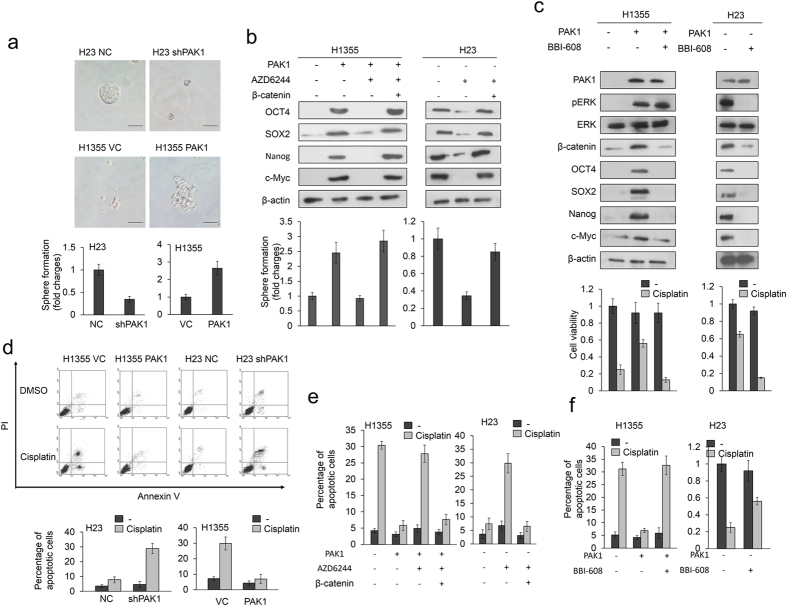
Figure 2. β-catenin-induced stemness is responsible for PAK1-mediated cisplatin resistance.
(a) Sphere formation abilities in H23 and PAK1-overexpressing H1355 cells were identified in a low-attachment plate by incubation in serum-free media for 1 week. The photographs show sphere colonies in H23 and PAK1-overexpressing H1355 cells (upper panel). The graphs show the average number of spheres in triplicate samples (lower panel). (b) H23 and PAK1-overexpressing H1355 cells were transfected with a β-catenin overexpression plasmid for 24 h, followed by treatment with the MEK/ERK inhibitor AZD6244 for 5 h. The cells were then incubated for 1 week in a low-attachment plate in serum-free media and evaluated for sphere formation abilities. Expression of the cancer stem cell markers OCT4, SOX2, Nanog, and cmyc was identified using western blotting. (c) H23 and PAK1-overexpressing H1355 cells were treated with a cell stemness inhibitor (10 μM BBI-608) for 5 h. The inhibitor was then removed and the cells were treated with 25 μM cisplatin for an additional 48 h. Cell viability was then evaluated by MTT assay. (d) H1355 and H23 cells were transfected with PAK1 expression vector and shPAK1 for 24 h. The cells were then treated with 0.1% DMSO or 25 μM cisplatin for 24 h and subjected to annexin-V and PI staining, followed by a flow cytometry analysis. The percentages of apoptotic cells in the annexin V+/PI- and the annexin-V+/PI+ populations were determined. (e) H23 and PAK1-overexpressing H1355 cells were transfected with β-catenin overexpression plasmid for 24 h, followed by treatment with AZD6244 for 5 h. The inhibitor was then removed and the cells were treated with cisplatin for an additional 48 h. (f) H23 and PAK1-overexpressing H1355 cells were treated with a cell stemness inhibitor (10 μM BBI-608) for 5 h. The inhibitor was then removed and the cells were treated with 25 μM cisplatin for an additional 48 h. Cell apoptosis was evaluated by flow cytometry. All experiments were performed three independent times. The mean values and the standard deviations are indicated as columns with error bars. The samples were derived from the same experiment and gels/blots were processed in parallel. Full-length blots are presented in Supplementary Figure S3.

