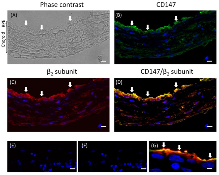
Figure 1.
Immunofluorescence of adult human eye in situ. (A) A phase-contrast image of the human eye section studied. The RPE layer and the choroid are indicated. The retina is already detached in the paraffin block. This section was co-stained for CD147 (B) and the β2 subunit of the Na+, K+-ATPase (C) using Alexa 488- and Alexa 594-conjugated donkey anti-rabbit and anti-mouse IgG secondary antibodies, respectively. The merged image showing co-localization at the apical domain is in (D). Panels (E,F) show similar sections treated only with the fluorescent secondary antibody (anti-rabbit and anti-mouse IgG, respectively) as negative controls. Panel (G) shows a higher-magnification image from a different field of the same preparation. All the preparations were counterstained with DAPI (blue). Arrows indicate the apical domain of RPE cells. Scale bars are 40 μm in (A–F) and 10 μm in (G).

