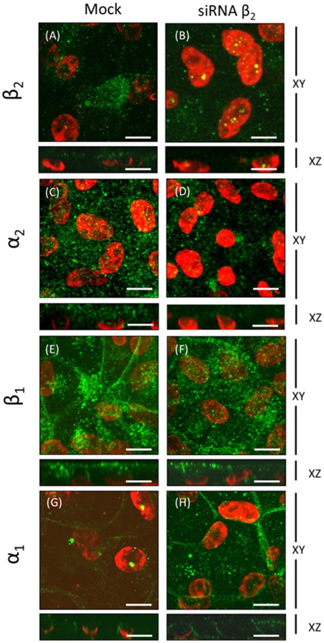
Figure 10.

Immunofluorescence analyses of ARPE-19 cells silenced with siRNA against the human β2 isoform. ARPE-19 cells cultured for 4 weeks were transfected with or without siRNA specific to the human β2 isoform, as shown in Figure 8. Confocal images of silenced (right panels) or not-silenced (left panels) cells immunostained for specific α and β isoforms are displayed. Apical expression of the β2 and α2 isoforms in mock-transfected cells is apparent in (A,C). Mislocalization and reductions in the fluorescence intensity of both β2 and α2 subunits are observed in silenced cells (B,D). Basolateral and apical expression of the β1 isoform (E) is generally maintained in β2-silenced cells. However, in the presented field, β1 has a noted apical distribution pattern (F). The mainly basolateral pattern of the α1 isoform (G,H) is maintained in silenced cells. Scale bar: 10 μm.
