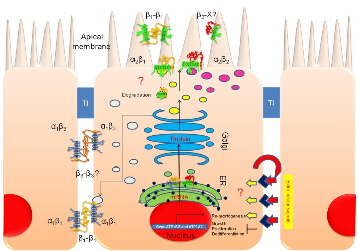
Figure 12.
The apical polarization of Na+, K+-ATPase in polarized ARPE-19 cells is regulated by the β2 subunit. An illustration of polarized ARPE-19 cells cultured for 4 weeks on permeable inserts in the presence of ITS is depicted; the cells are relatively tall, express RPE markers and form adjacent tight junctions (TJs). Our model proposes that extracellular signals trigger transduction pathways that activate re-morphogenesis. In non-polarized ARPE-19 cells, a basolateral targeting mechanism carries α1β1 and α1β3 dimers to the lateral membrane (gray vesicles). The β1-β1 (and perhaps β3-β3) trans-interaction between neighboring cells stabilizes and retains these dimers at the lateral membrane domain for housekeeping. Upon the triggering of re-morphogenesis, a concurrent apical targeting mechanism is activated by the association of the α2 and β2 subunits. α2β2 is delivered to the apical domain (magenta vesicles). In cultures, this complex probably does not stabilize at the apical plasma membrane but accumulates in a sub-apical compartment. In situ, the pump is inserted in the apical membrane domain, where it is probably stabilized by heterotypic trans interaction(s) of the β2 subunit with adhesion proteins on the outer segments of the photoreceptor (β2-X).

