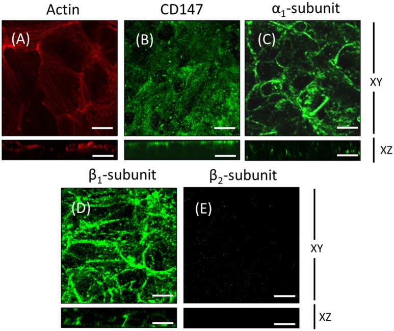
Figure 2.
ARPE-19 cells cultured on transwell inserts for 4 weeks are not completely polarized. ARPE-19 cells were cultured up to 4 weeks on transwell inserts covered with laminin. The immunofluorescence image in (A) shows actin localization using rhodamine phalloidin. The cells are flat with stress fibers and very little circumferential actin microfilament bundles. The expression of CD147, a RPE marker, was detected using a specific antibody in the apical membrane domain (B). The expression of Na+, K+-ATPase using anti-α1 (C) and anti-β1 antibodies (D) was observed mostly at the basolateral membrane. Immunofluorescence detection with anti-human β2 antibody revealed a very weak signal (E). Scale bar: 10 μm.

