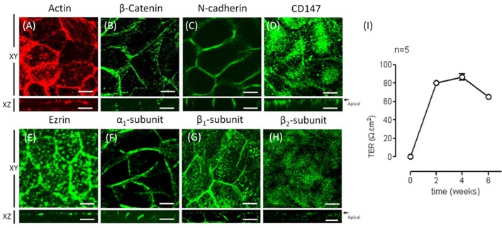
Figure 3.
Re-morphogenesis of ARPE-19 cells treated with ITS. (A) The photomicrographs show ARPE-19 cells cultured for 4 weeks with ITS and stained for actin with Texas Red-X phalloidin. Note the tight intercellular contact, epithelial-like cellular shape, and predominantly peripheral cortical actin staining. Confocal immunofluorescence images of the same culture conditions showing β-catenin (B) and N-cadherin (C) at the lateral membrane, CD147 (D) mostly in the apical domain and ezrin (E) in the apical domain. (F) The immunofluorescence image shows immunostaining of the α1 subunit of Na+, K+-ATPase at the lateral membrane and more precisely, as observed in the XZ image, at the cell-cell contacts. (G) The β1 subunit is observed at the cell border and at the apical domain; the XZ image confirms both basolateral and apical staining. (H) The β2 subunit is observed mainly at the apical domain; the XZ image confirms apical staining. Scale bar: 10 μm. (I) Quantitative analysis of the transepithelial resistance (TER) of ARPE-19 cells during re-morphogenesis is depicted. Note a stable TER of ~80 Ω cm2 between the second and fourth week in culture.

