Figure 4.

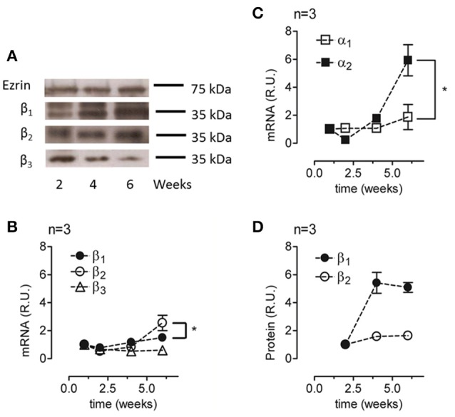
Analyses of the relative amounts of α and β isoforms in ARPE-19 cells during re-morphogenesis. (A) Western blot analysis of the lysates of ARPE-19 cells cultured for the indicated time under conditions established for re-morphogenesis. The upper part of the blot was probed with an antibody against ezrin (as a loading control), whereas the lower part was probed with antibodies against the β1, β2, and β3 subunits. Western blot analyses of all samples were conducted on the same day. A non-relevant lane between 2 and 4 weeks in the ezrin blot was eliminated; therefore, it appears to be discontinued. The blots represent three different experiments. Total mRNA was extracted at weeks 1, 2, 4, and 6 of culture and analyzed via qPCR. The relative mRNA levels of the β1, β2, and β3 subunits (B) and of the α1 and α2 subunits (C) are illustrated. The amount of mRNA was normalized to that detected in the first week. (D) Quantification of β1 and β2 subunit expression via densitometry in three independent experiments normalized to the loading control. Densitometry results were also corrected for the film light level whenever the background level was different. Error bars represent the mean ± SEM of three independent experiments. Significant changes are indicated by an asterisk (P < 0.05, non-parametric t-test).
