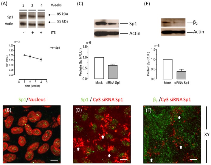
Figure 5.
The transcription factor Sp1 is involved in β2 subunit regulation in ARPE-19 cells. (A) Western blot analysis showing the expression of Sp1 over 4 weeks of culturing (upper panel). Actin was used as a loading control in all Western blots shown in this figure. Quantitative analysis of Sp1 normalized to the loading control is illustrated in the lower panel. (B) Immunofluorescent staining of Sp1 in ARPE-19 cells is shown in green. Counterstaining of nuclei is shown in red. Merged image shows the nuclear localization of Sp1 in ARPE-19 cells cultured for 4 weeks in the presence of ITS. Scale bar: 20 μm. (C) Representative Western blot and quantitative analyses of six independent experiments conducted on ARPE-19 cells treated with Sp1 siRNA are shown. Control cells were transfected without siRNA (Mock). Error bars represent the mean ± SEM. (D) Immunofluorescence image of ARPE-19 cells incubated with Cy3-siRNAs to silence Sp1 (red) and immunostained for Sp1 expression (green). White arrows indicate siRNA-transfected cells (identified by red fluorescence) that did not express Sp1 in the nucleus. Scale bar: 20 μm. (E) Representative Western blot and quantitative results of the immunodetection of the β2 subunit in six independent experiments of Sp1 knockdown in cells (60%). (F) Immunofluorescence image of Sp1-silenced ARPE-19 cells stained for β2 subunit expression.

