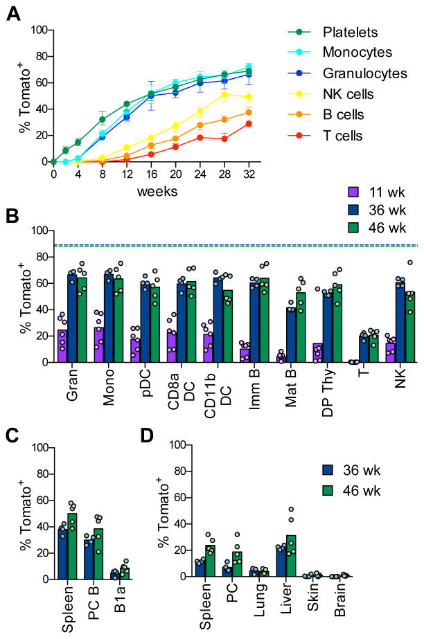
Fig. 6. Labeled HSCs provide a major contribution to mature hematopoietic cells.
A. The fraction of labeled Tom+ cells in peripheral blood cells of Pdzk1ip1-CreER R26Tom/Tom reporter mice at the indicated time points after tamoxifen administration. (median ± range of 4 animals, representative of two experiments).
B–D. The fraction of labeled Tom+ cells in mature cell types of reporter mice. Shown are the median (bars) and values from individual animals (circles) at the indicated time points after tamoxifen treatment.
B. Major immune cell types in the spleen or thymus. The median fractions of Tom+ HSCs at the respective time points are indicated by dashed lines.
C. Conventional B cells in the spleen or peritoneal cavity (PC) and B-1a cells in PC.
D. Resident macrophages from the indicated tissues.
See also Fig. S6.

