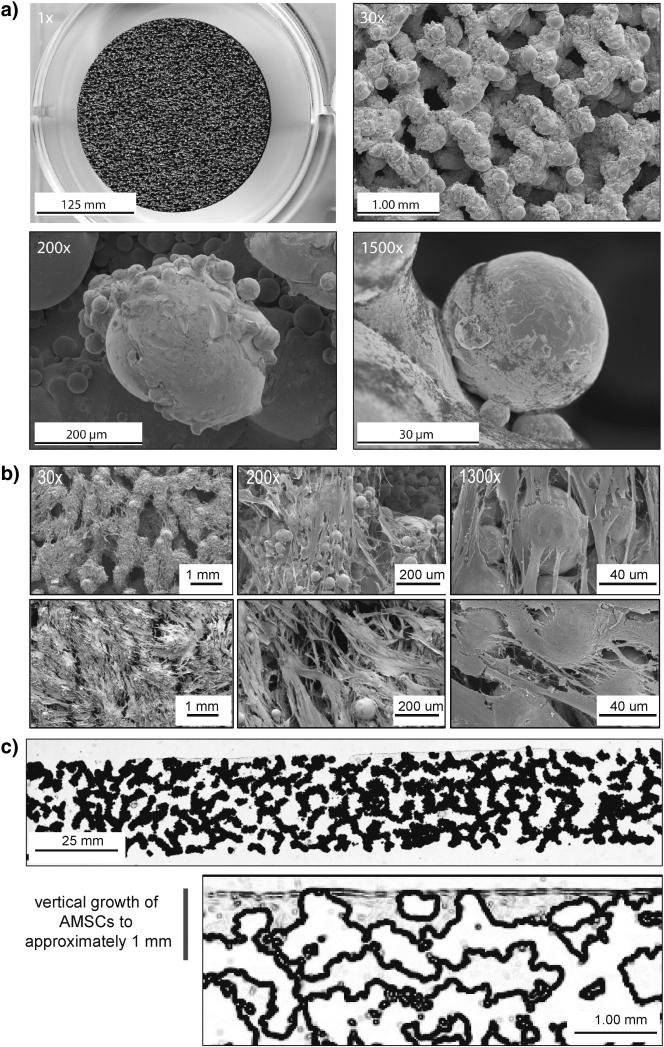
Figure 1.
(a) Scanning electron microscopy images of porous structured titanium (TiAl6V4) at 1x, 30x, 200x, and 1500x. (b) SEM images after 7 days in culture at 30x, 200x, and 1300x. (c) Histology slide of Sanderson's rapid bone stain demonstrating vertical penetration into pores of ps-Ti to an approximate depth of 1 mm. Samples were taken from donor AMSC-258 after 14 days. Close-up image of Sanderson's Rapid Bone Stain (Collagen = Grey).

