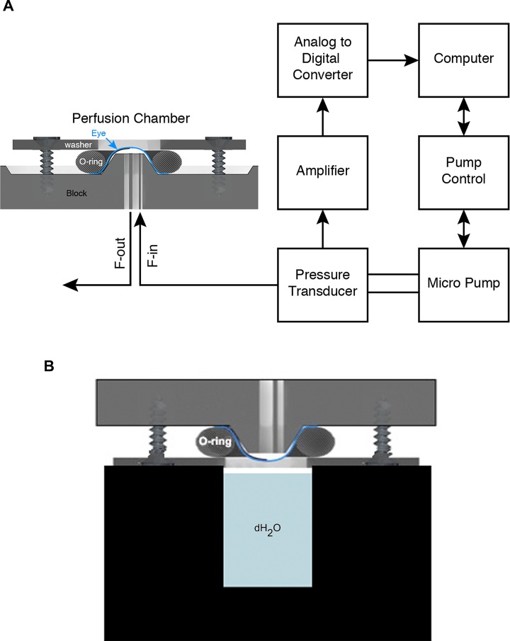
Figure 2.
Schematic of system control. (A) The eye is sealed onto the perfusion device and fluid (saline, drugs, and tracers) is introduced or drained through the two ports (F-in, F-out). F-out is closed except when flushing. The F-in port is connected to a pressure transducer, which in turn is connected to a micropump. The pressure transducer monitors IOP constantly and supplies this information to the computer, which control the pump controller and pump. The control algorithm maintains pressures at the desired values and durations according to the preset experimental plan. (B) Schematic showing a mounted eye in a humidifying chamber that is maintained at 37°C (not to scale).

