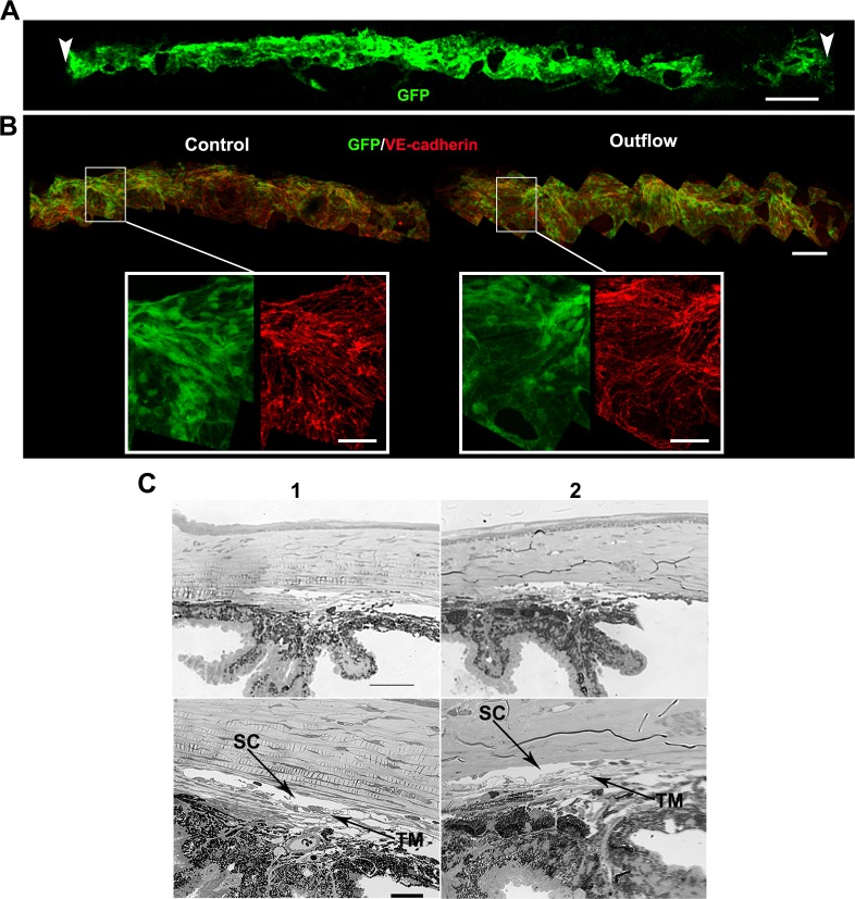
Figure 5.
The drainage structures remain intact during mounting and perfusion. (A) Whole mount of a Prox1-GFP eye after an outflow experiment, showing the entire extent of the limbus and SC (green) subjected to flow. Schlemm's canal over this entire length has a normal healthy morphology. Vascular endothelial–cadherin staining (not shown) was present in area low in Prox1-GFP at the right. Arrowheads indicate points where the O-ring contacted the limbus when the eye was mounted on the frustum. Typically approximately 45% of the limbus is subjected to flow (see Supplementary Methods). (B) Higher magnification images of a freshly dissected control eye and an eye after outflow. The general morphology (GFP, green) and cellular junctions (VE-cadherin, red) of SC are indistinguishable between these eyes. This indicates that the inner wall SC endothelium is intact. The GFP level in eyes subjected to outflow is always within the range of variability in unperfused control Prox1-GFP eyes. Retention of GFP signal after outflow is a good indicator that SC cells are healthy. (It is well established that compromised cell membrane integrity results in leakage of cytoplasmic GFP and reduction in green fluorescence.) n = 6 eyes each for control and outflow cohorts. Scales bars: (A) 300 μm; (B) top 100 μm; insets 30 μm. (C) Plastic sections of a control (1) and perfused (2) eye showing the drainage structures. n = 3 eyes each for control and outflow cohorts. Images at lower magnification show the angle in the context of the iris and cornea (top). Higher magnification images (bottom) allow for visualization of the open SC and TM. Scale bars: Top, 50 μm; Bottom, 20 μm.

