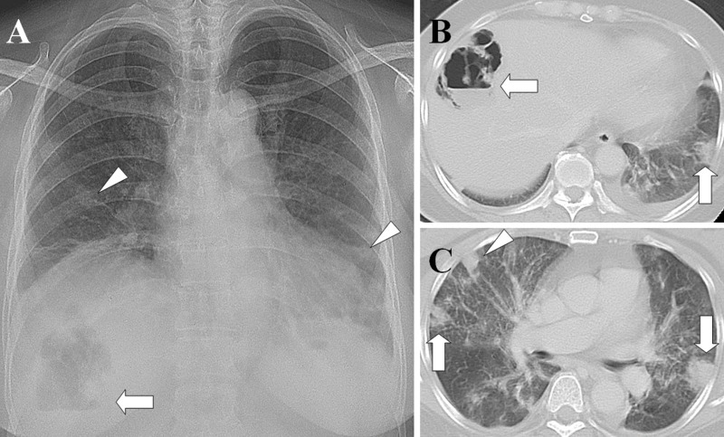
Figure 1.
Liver abscess with septic pulmonary emboli. (A) A chest radiograph shows an air-fluid level in the right upper abdomen (arrow) and multiple nodular opacities in the bilateral lungs (arrowheads). (B) A CT scan shows a gas-forming liver abscess (arrow) and a peripheral wedge-shaped opacity abutting the adjacent pleura (arrow). (C) A lung window of a cross-sectional CT scan shows two peripheral wedge-shaped opacities abutting the adjacent pleura (arrows) and a peripheral nodule with a feeding vessel (arrowhead). The patient was a 61-year-old diabetic woman whose blood and aspirate abscess cultures were positive for Klebsiella pneumoniae.

