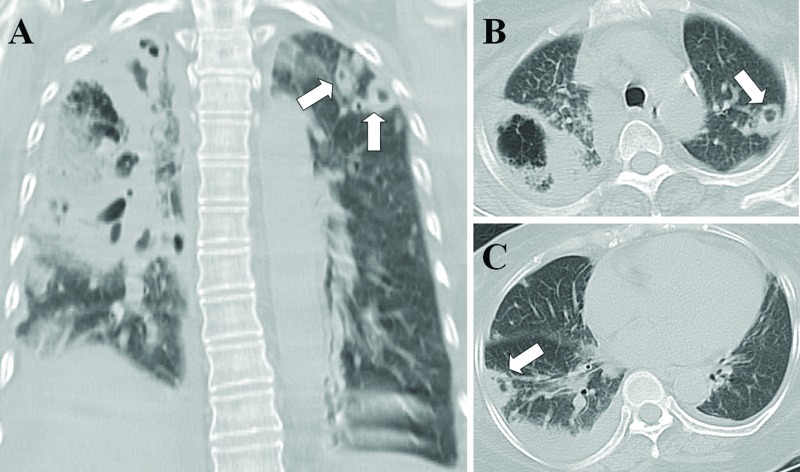
Figure 2.
Necrotizing pneumonia with septic pulmonary emboli. (A) A lung window of a coronal-sectional CT scan shows necrotizing pneumonia in the right lung. Multiple different sizes of nodules with cavities in the left upper lobe (arrows), suggestive of septic pulmonary emboli, are observed. (B) A lung window of a cross-sectional CT scan shows necrotizing pneumonia in the right upper lobe and a cavitary nodule in the left upper lobe (arrows). (C) A peripheral wedge-shaped opacity abutting the adjacent pleura in the right lower lobe (arrow) and pleural effusion are seen. The patient was a 62-year-old diabetic woman whose blood and sputum cultures were positive for methicillin-resistant Staphylococcus aureus.

