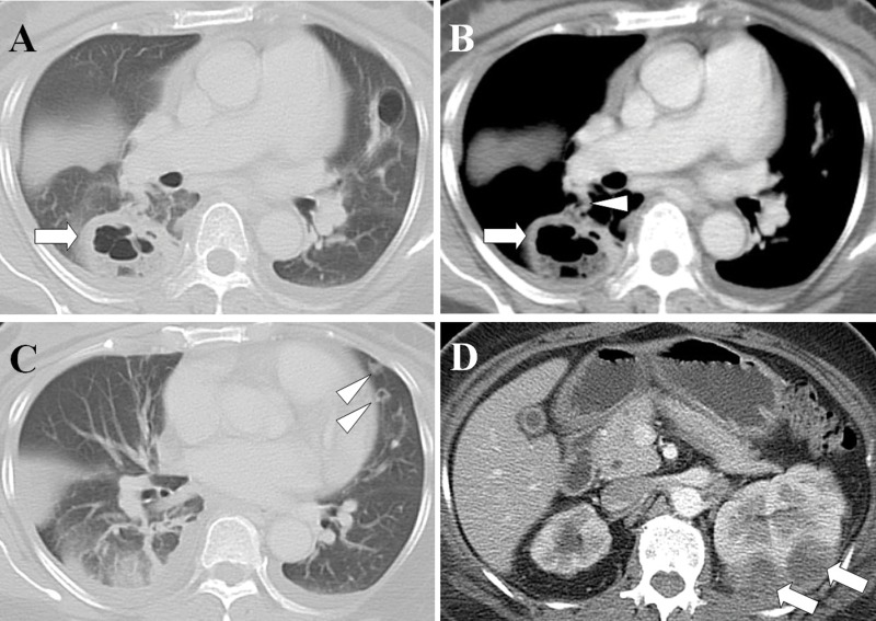
Figure 4.
Renal abscesses with septic pulmonary emboli. (A) A lung window of a cross-sectional CT scan shows a lung abscess with a diameter of 4.5 cm in the right lower lobe (arrow). (B) A contrast-enhanced CT scan (mediastinum window) in the same image plane shows a lung abscess (arrow) with a feeding vessel sign (arrowhead). (C) A lung window of a cross-sectional CT scan shows two nodules with cavities in the left upper lobe (arrowheads). (D) An abdominal CT scan shows left renal abscesses (arrows). The patient was a 52-year-old woman whose blood cultures were positive for Escherichia coli.

