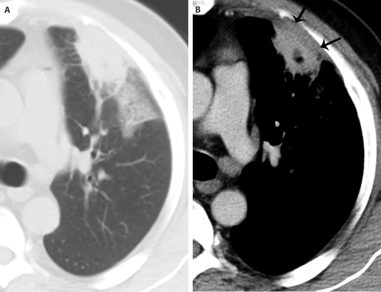
Figure 2.
An inflammatory patch in a 48-year-old man with cough, expectoration and chest pain. There is a subpleural patch in the superior lobe of the left lung. Its interface is ill-defined, and ground glass opacity can be detected in the peripheral lung field (A). The basement of the lesion is broad. On the enhanced image, there is a low-density area with a clear boundary in it, which indicates necrosis. Significant pleural thickening (arrows) can be seen (B).

