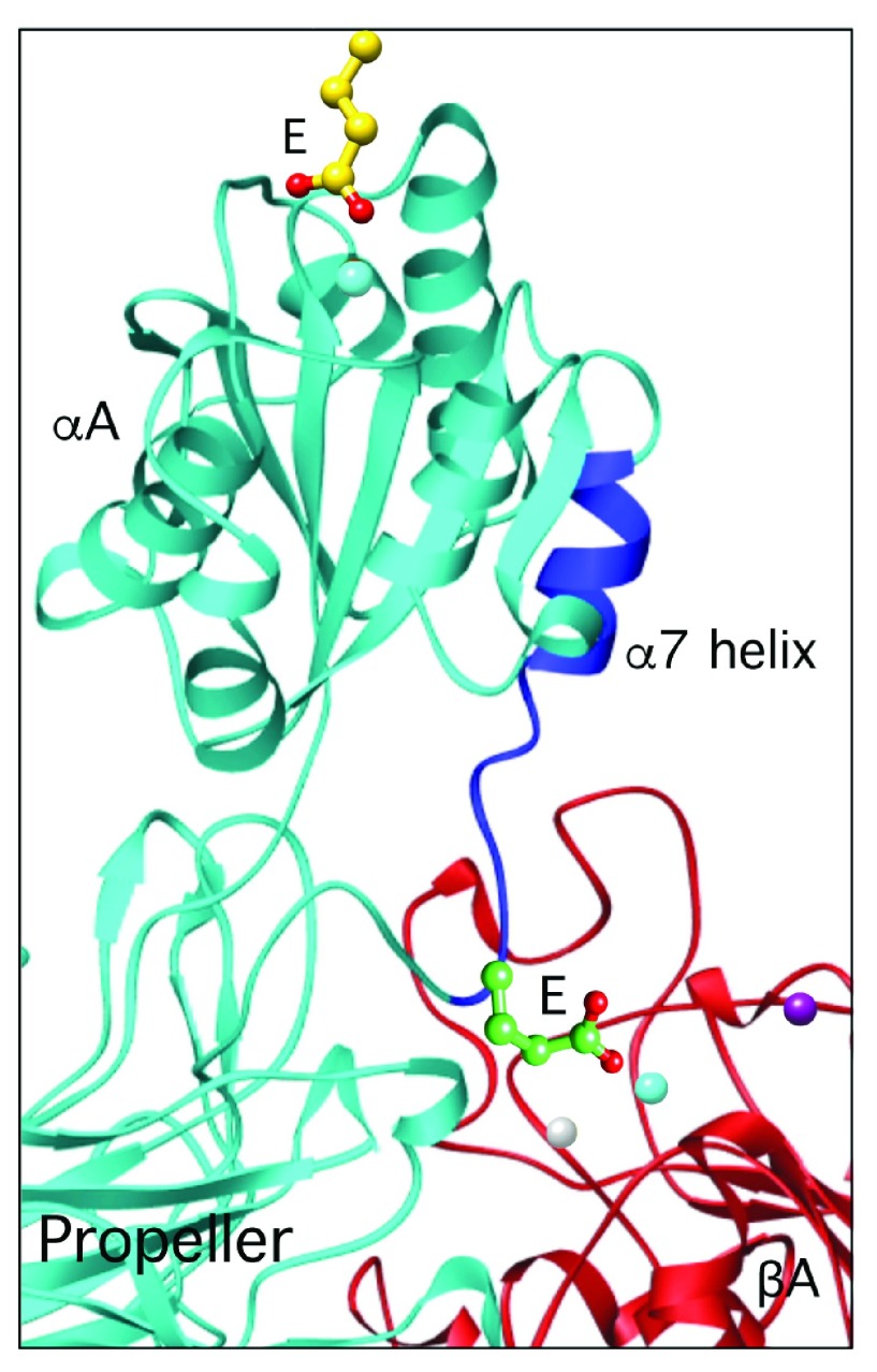
Figure 4. The ligand-relay model.
The downward movement of the c-terminal α7 helix (dark blue) triggered by ligand binding to αA allows an invariant glutamate (E) at the bottom of the α7 helix to reach and ligate the βA metal-ion-dependent adhesion site (MIDAS) ion (cyan), thus relaying the ligand occupancy state of αA to βA.

