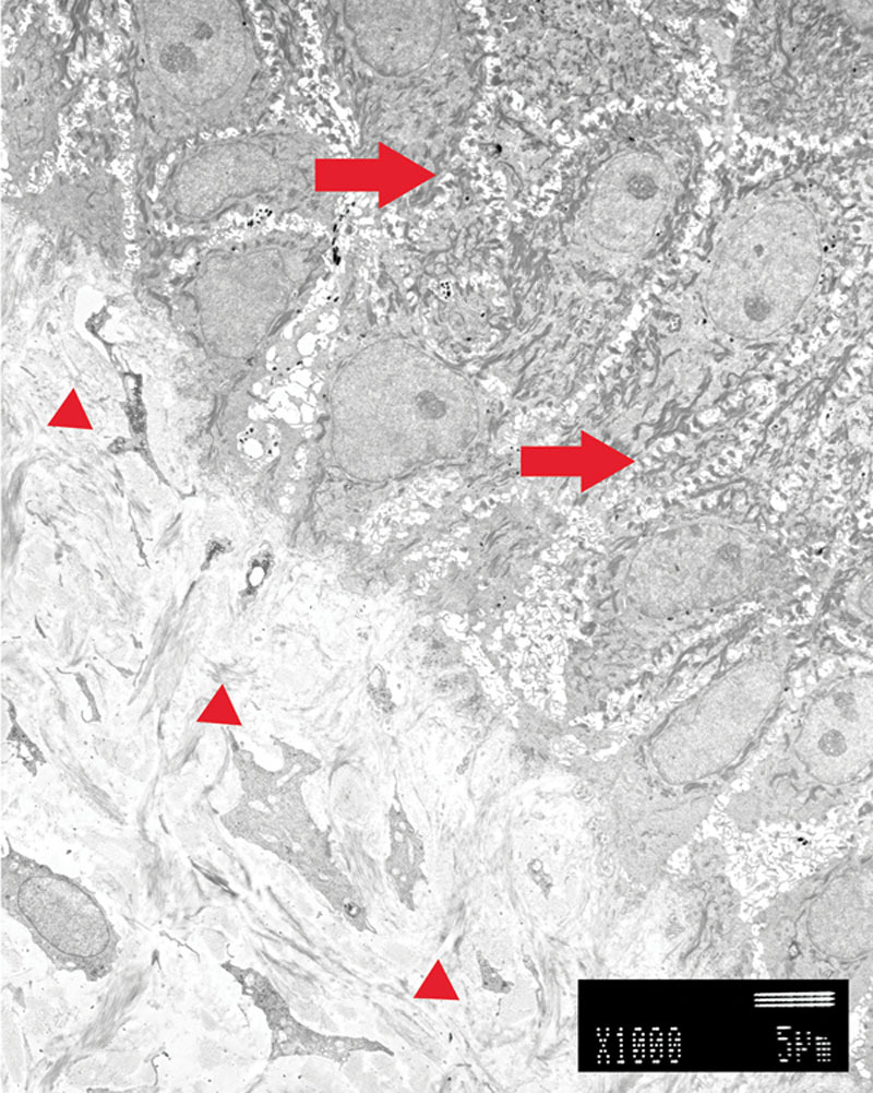
Fig. 3.

Transmission electron microscope findings of autograft. Keratinocytes were located in the upper part. Dermal layer was located in the lower part. We recognized a lot of thick desmosomes between the keratinocytes (right arrows). Basement membrane was observed in the lowest layer of keratinocytes. In the dermal layer, various thickness collagen fibers were oriented in various directions (small arrowheads).
