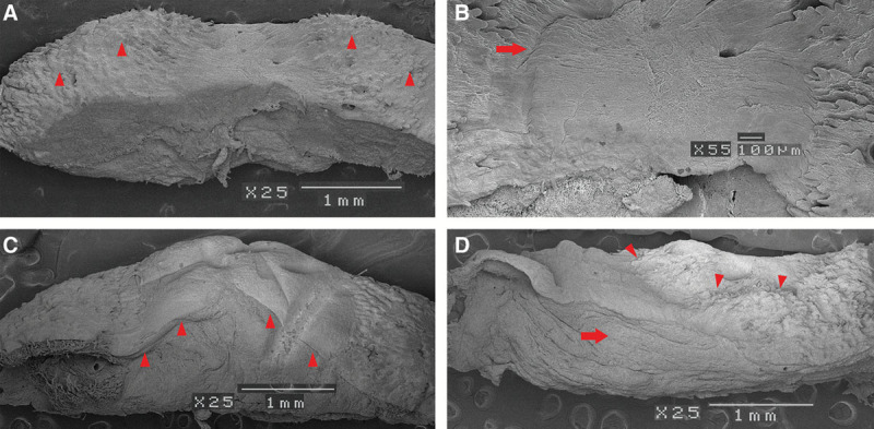
Fig. 6.

A, Scanning electron microscope findings. Two weeks after grafting. Because the epithelium disappeared, we were able to observe the structure of the dermis layer. At both ends, we recognized papillary dermis (small arrowheads). The uneven surface formed a rete ridge. B, Two weeks after grafting. Both ends of the papillary dermis were extended toward the center of the top of the dermal-like tissue (right arrow). C, Four weeks after grafting. New network of collagen fibers continuing from the papillary dermis were cross-linked from both ends and covered all dermal-like tissue (small arrowheads). D, Six weeks after grafting. On the upper surface of the specimen, irregularities similar to papillary dermis were observed (small arrowheads). The structure of the dermis-like tissue was almost the same as autodermis (right arrow).
