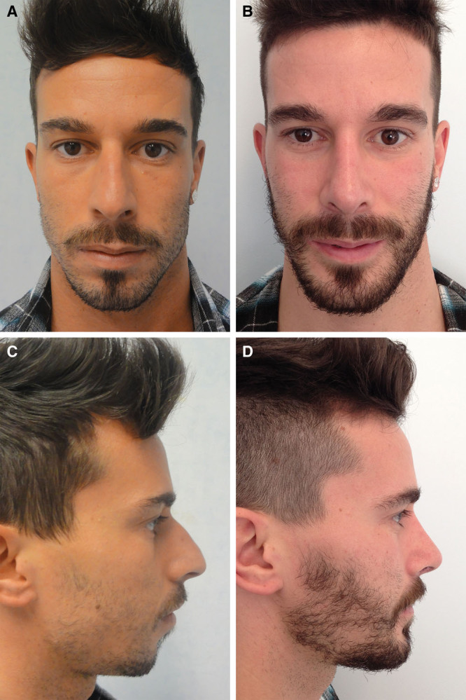
Fig. 3.

Case 1: Deviation of the nasal spine. Preoperative (A, C) and postoperative (B, D) photographs on frontal and profile views. Correction of the defects involved osteocartilaginous hump excision, osteotomies, genioplasty, fracturing, and median repositioning of the nasal spine combined with suturing to the periosteum of the upper maxillary bone.
