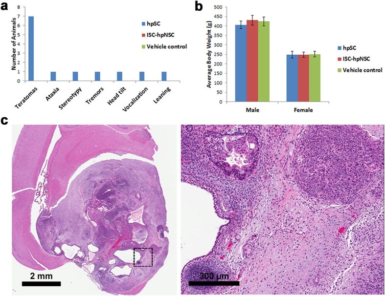
Figure 5. Tumorigenicity and biodistribution study of ISC-hpNSC.
(a) Number of animals with teratomas, ataxia, stereotypy, tremors, head tilt, vocalization, and leaning in the hpSC, ISC-hpNSC, and vehicle control groups. (b) Average body weight of the hpSC, ISC-hpNSC, and vehicle control groups with no statistically significant differences between the groups. (c) Representative image of a teratoma found in the brain of a rat injected with undifferentiated hpSC with disorderly growth with red blood cells (red area) and numerous clear spaces. The black dotted square represents the area where the higher magnification image on the right was taken, showing the presence of cartilage (bottom left corner), mucous-producing cells (top left) and nervous tissue, representing disorderly endodermal and ectodermal differentiation as assessed by an experienced board certified pathologist.

