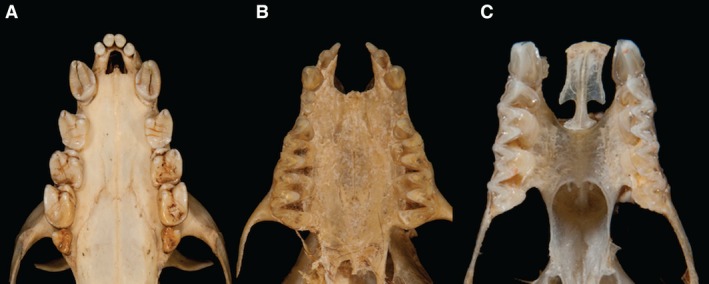
Figure 1.

Patterns of variation in the chiropteran premaxilla. (A) Acerodon jubatus (Pteropodidae). The anterior dento‐alveolar arch is intact with no bony cleft. The bony margins of the incisive foramen are reduced, resulting in an enlarged incisive foramen. (B) Myotis myotis (Vespertilionidae). The anterior dento‐alveolar arch is interrupted by a U‐shaped, midline bony cleft between the two premaxillae. Each premaxilla bears incisors and is fused laterally to the maxilla. The cleft extends posteriorly to the anterior margin of the palatal shelves of the maxillae and is continuous with the incisive foramina. (C) Rhinolophus ferrumequinum (Rhinolophidae). The anterior dento‐alveolar arch is interrupted by bilateral paramedian clefts between the premaxillae (which articulate across the midline via a sutural joint and bear diminutive incisors) and the maxillae. The clefts extend posteriorly to the anterior margin of the palatal shelves of the maxillae and are continuous with the incisive foramina. (Photographs: Phil Myers, Museum of Zoology, University of Michigan‐Ann Arbor, www.animaldiversity.org. Creative Commons Attribution‐Noncommercial‐Share Alike 3.0 Unported License).
