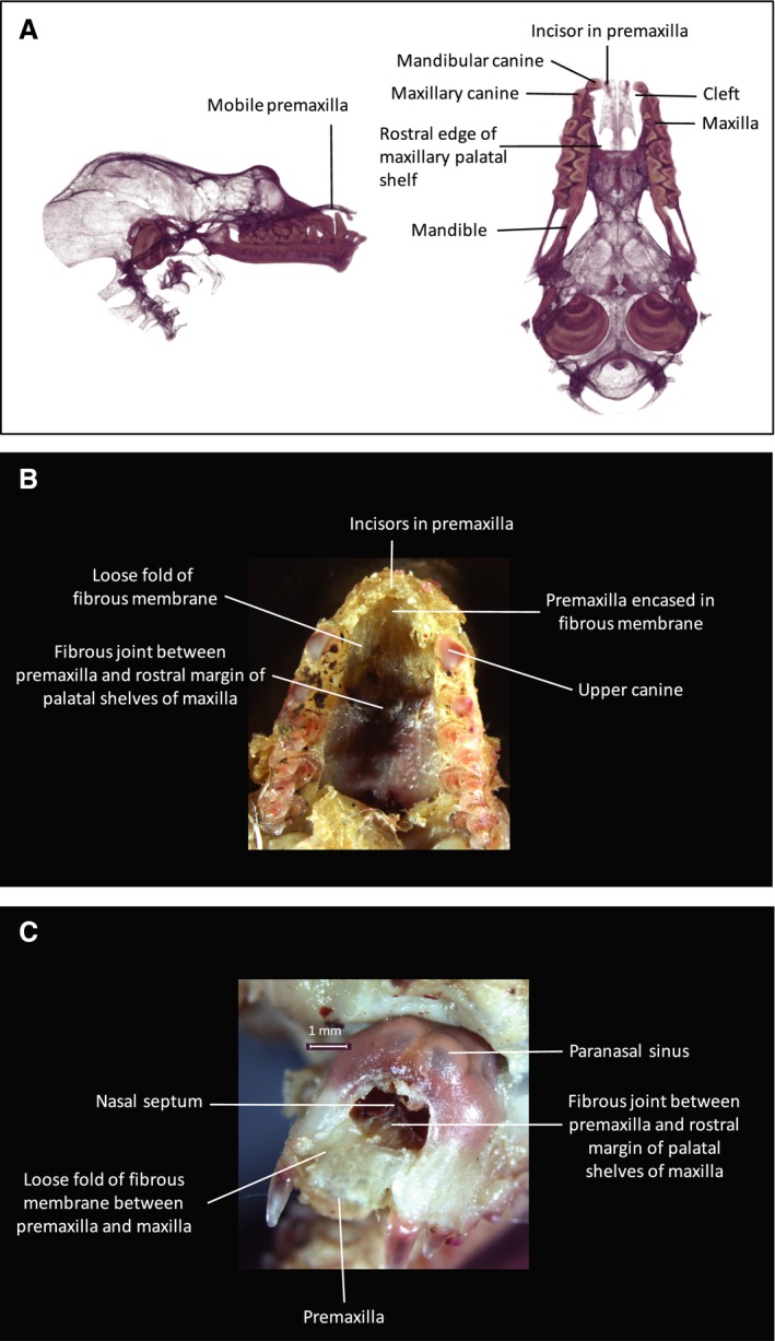
Figure 10.

Cranial anatomy of Rhinolophus euryale. (A) 3D μCT inferior and lateral views of cranial skeleton with body of mandible digitally subtracted. Cusps of mandibular teeth are seen occluding medial to the maxillary dental arch. Bilateral paramedian bony clefts are present in the dento‐alveolar arch between the premaxilla and the maxilla. The clefts extend posteriorly to the anterior margin of the maxillary palatal shelves, to which the posterior extremities of the premaxillae are attached via a midline sutural joint. The notched posterolateral margin of the premaxilla represents the site of the incisive foramen on either side. The premaxillae are united in the midline by an unfused sutural joint. The premaxilla is seen to float superior to the occlusal plane, hinging on the midline articulation with the anterior margin of the maxillary palatine shelves. (B) Inferior view of dissection specimen to show anterior palate. (C) Anterior view of dissection specimen to show facial rostrum. The bony cleft is completely filled in by a robust, fibrous membrane that is continuous with the periosteum of the maxillary margins of the cleft and splits to enclose the premaxillae. The membrane is loose in the space between the maxilla and the premaxilla, allowing the premaxilla to move freely in a sagittal plane as it hinges about its posterior sutural joint with the palatal shelf of the maxilla. The nose‐leaf cartilages are attached to the superior surface of the premaxillae and are open superiorly. Thus the mobile premaxilla forms a mobile base to the nose‐leaf, anterior to the nasal cavity. Bullae of the maxillary paranasal sinuses are visible in (C), superior to the anterior bony margin of the nasal cavity.
