Figure 11.

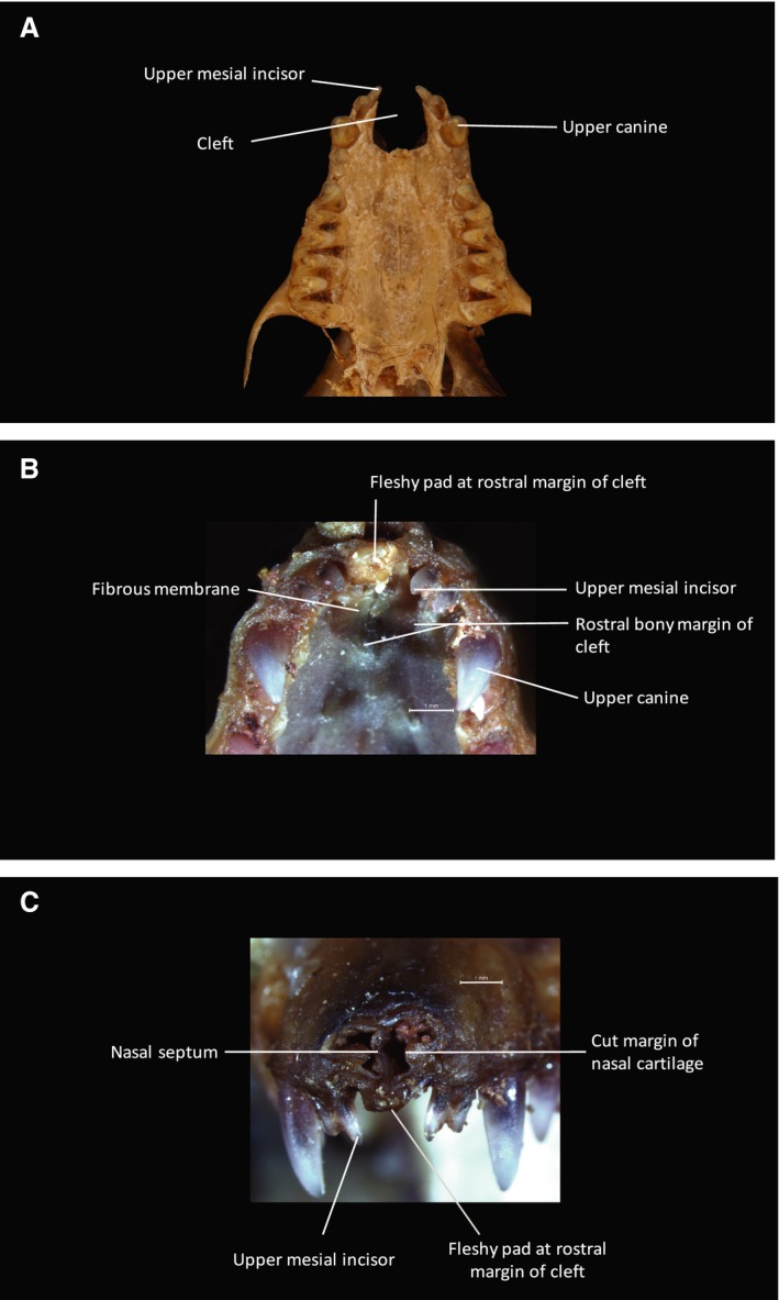
Cranial anatomy of Myotis. (A) Inferior view of cranial skeleton of Myotis myotis to show palate. A U‐shaped bony cleft is present between the two premaxillae. Each premaxilla, bearing two conical incisors mesial to the large and pointed maxillary canine, is fused to the maxilla. The cleft is bounded posteriorly by the anterior free bony margin of the palatal shelves. (B) Inferior view of dissection specimen (Myotis blythii) to show anterior palate. (C) Anterior view of dissection specimen to show facial rostrum. As in Plecotus (Fig. 9) a robust, fibrous membrane, continuous with the periosteum at the margins, fills in the U‐shaped cleft visible in (A). The anterior free margin of this membrane terminates as a fibrous condensation. The nasal septum (visible in C) attaches to this membrane and the fibrous condensation in the midline. (Photograph (A) Phil Myers, Museum of Zoology, University of Michigan‐Ann Arbor, www.animaldiversity.org. Creative Commons Attribution‐Noncommercial‐Share Alike 3.0 Unported License).
