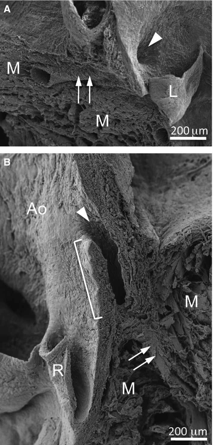
Figure 3.

The normal and intramural courses of the coronary artery stems. Scanning electron micrographs of the left (A) and right (B) aortic sinus and adjacent myocardium of a Balb/c (A) and a C57BL/6 mouse (B). (A) The left coronary ostium (arrowhead) is located at the level where the myocardium contacted the aortic root, and the coronary artery stem (arrows) runs intramyocardially from its origin. (B) The right coronary ostium (arrowhead) shows a slit‐like shape. The bracket indicates the portion of the coronary artery stem with an intramural course. The arrows point to the intramyocardial portion of the coronary artery. Ao, aorta; L, left leaflet; M, myocardium; R, right leaflet.
