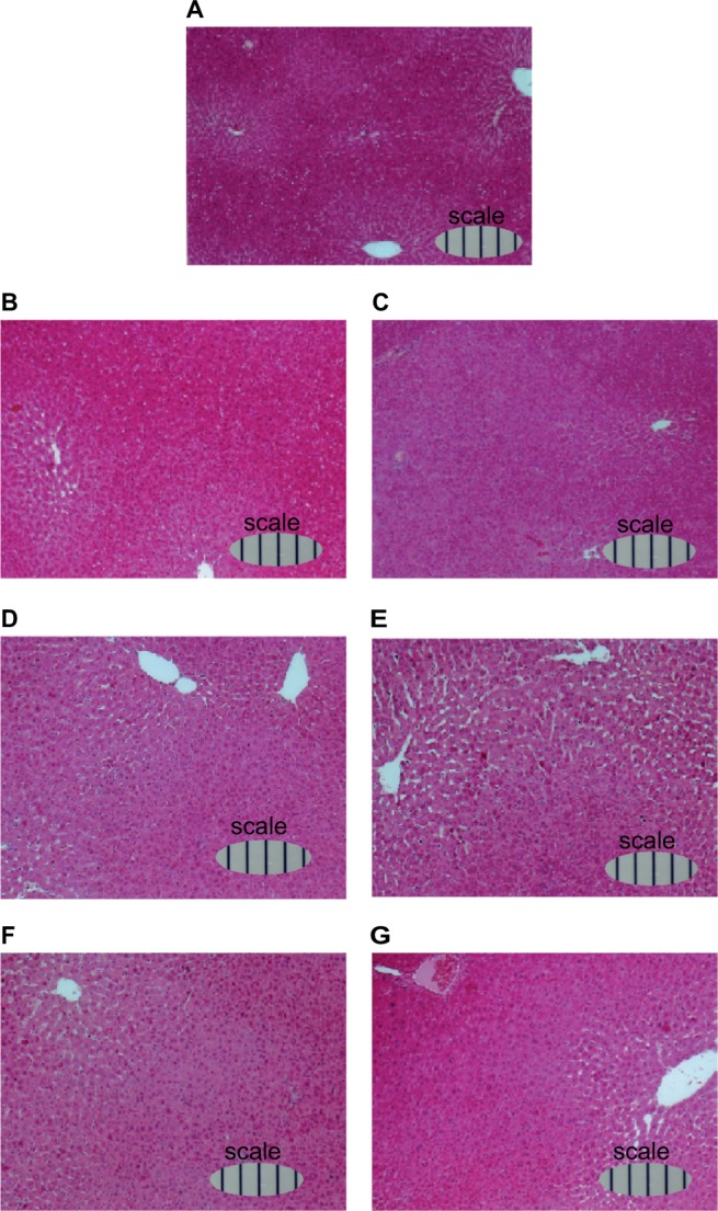
Figure 8.

Light microphotographs of H&E-stained liver sections of rats during liver regeneration after 70% PH (×10).
Notes: Rats were sacrificed at (A) day 0, (B) day 1, (C) day 2, (D) day 3, (E) day 4, (F) day 5, and (G) day 6 after 70% PH, the livers were immediately removed, and weighed. Tissue samples were routinely fixed in 4% neutral buffered formalin for several days, dehydrated, paraffin-embedded, and sectioned at 5 µm thickness. Following deparaffinization in xylene, sections were dehydrated, stained with H&E, and mounted on glass slides for pathological assessment. Scale bars show in 100 µm.
Abbreviations: H&E, hematoxylin and eosin; PH, partial hepatectomy.
