
Figure 2.
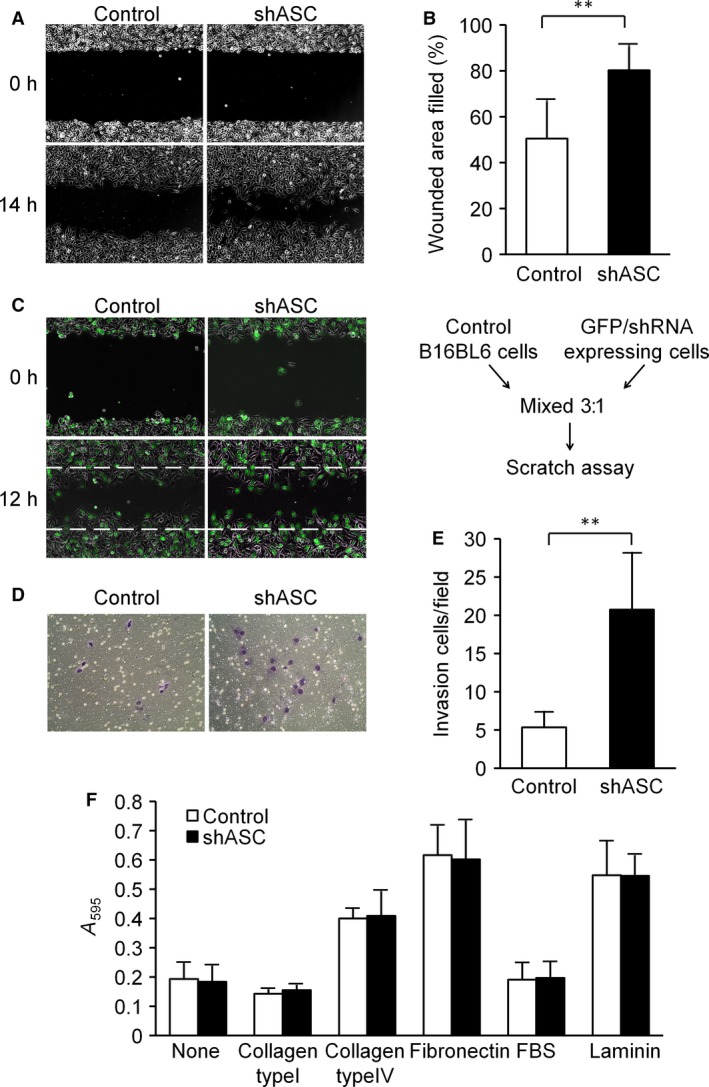
ASC silencing significantly enhanced the motility of B16BL6 cells. (A and B) Scratch assay. (A) Representative photographs taken immediately (0 h) and 14 h after wounding. (B) The wounded areas were measured by ImageJ software, and the percent of wound closure was calculated for each area. Results are expressed as the mean (n = 6), and bars indicate SD. **P < 0.01. (C) Scratch assay with GFP‐labeled cells. Parental and GFP/shRNA‐expressing cells were mixed at a ratio of 3: 1 and then seeded in the same manner as in experiment A. Migration progress was photographed immediately (0 h) and 12 h after wounding under a fluorescent‐inverted microscope. Representative results are shown. (D and E) Transwell invasion assay. (D) Representative photographs of invading cells. (E) Invading cells were counted in five randomly chosen fields in each wells. Results are expressed as the mean of triplicate counts (n = 15) and bars indicate SD. **P < 0.01. F. ECM adhesion assay. After pre‐coating 96‐well plates with the indicated ECM molecules, adhesion assays were performed as described in Materials and Methods. Results are expressed as the mean (n = 4) and bars indicate SD.