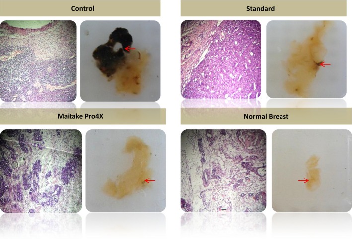
Figure 4.

Histology of mammary tissues from each treatment group with or without Maitake. The picture indicated as normal breast corresponded to breast tissues without tumor developed after Maitake Pro4X treatment. Right picture shows the paraffin block of breast tissues, the arrow indicate the amplified region and left picture shows the corresponding microscopy breast histology.
