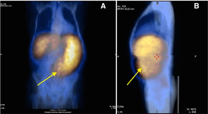
Figure 3.

111In‐Cl3 single‐photon emission tomography/computed tomography imaging in one patient with very advanced primary myelofibrosis: (A) front view and (B) side view showing very intense uptake of the radiotracer in the spleen (yellow arrow) and almost no fixation in the backbone and other structures of the axial skeleton.
