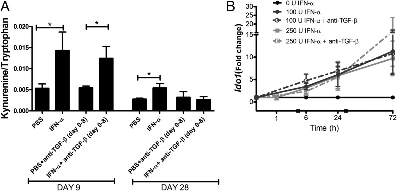
FIGURE 5.
IFN-α–induced activation of IDO1 in the sensitization phase of AIA and in vitro occurs independently of TGF-β. Female mice were immunized days 1 and 7 with mBSA with or without IFN-α treatment, with or without anti–TGF-β Abs administered i.p. days 1–8 as in Fig. 1 and as described for AIA in Materials and Methods. (A) Sera were collected at days 9 and 28 and analyzed for Trp and Kyn concentration by HPLC. Data are expressed as the ratio of serum levels of Kyn to Trp, n ≥ 7. Comparisons between groups were made by Mann–Whitney U test. (B) Splenocytes isolated day 10 from mBSA-sensitized mice were restimulated ex vivo with 50 μg/ml mBSA with or without 100 U/ml IFN-α, with or without 40 μg/ml anti–TGF-β. Data are expressed as fold change normalized to the reference gene Actb and Ido1 expression in mBSA-stimulated cultured cells from the same mice, n = 5. Paired Student t test was used to evaluate differences between treatments. *p < 0.05.

