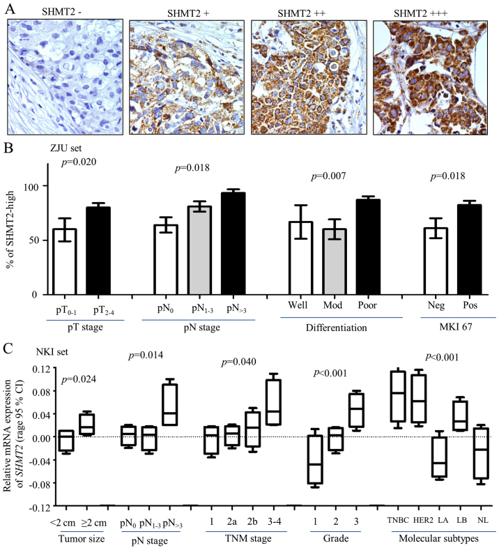
Figure 2.
Clinical relevance of SHMT2 in breast cancer. Protein expression levels of SHMT2 and other biomarkers were determined using immunohistochemistry (IHC) staining. (A) Representative IHC images stained with SHMT2 (magnification, ×200). (B) SHMT2 protein expression levels associated with pathological tumor and lymph node stage (pT and pN stages), poor differentiation and MKI-67 in ZJU set. Here, pT is pathological tumor stage; and the number represent tumor stage. pN is pathological lymph node stage; the number indicates how many lymph nodes involved. pN>3 is >3 lymph nodes were involved. Mod, moderate differentiation; Neg, negative; Pos, positive. (C) ANOVA analysis result for SHMT2 mRNA expression levels and tumor size, pN and TNM stages, Elson grade, and intrinsic molecular subtypes of breast cancer. TNBC, basal-like breast cancer; HER2, Her2-positive, LA, luminal A; LB, luminal B; NL, normal breast-like. The p-values represent overall ANOVA analysis results.

