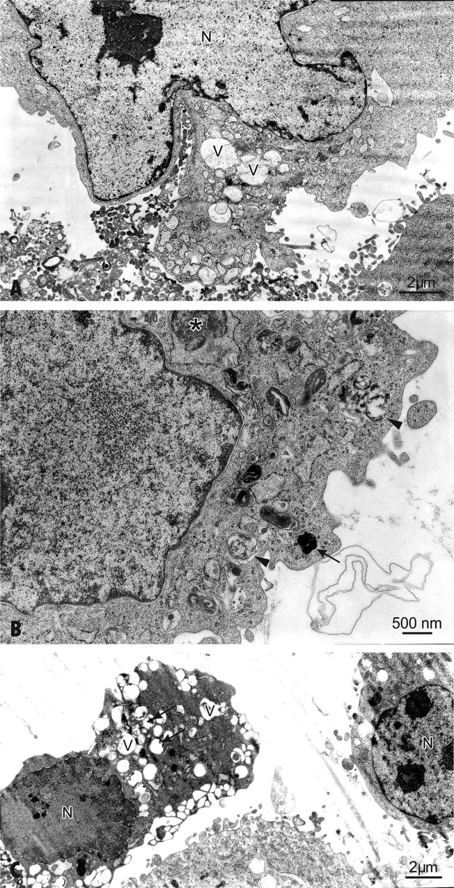
Fig 3. The ultrastructural alterations of hFOB 1.19 cells after exposure to 30 or 60 μg/mL AgNPs for 48 h.
(A) Cells show ultrastructural changes and formation of multiple blebs. Enlarged nucleus (N), pushing the cytoplasm towards the periphery. (B) The cell exposed to 30 μg/mL AgNPs for 48 h contains double-membraned autophagic vacuoles—autophagolysosomes with engulfed organelles displaying degenerative changes (arrowheads). Autophagosome (asterisk) and lysosome (arrow) are indicated. Note condensed mitochondria and disorganization of the inner membrane system. (C) Apoptosis in cells exposed to 60 μg/mL AgNPs for 48 h. A decrease in cell volume and chromatin condensation show induction of apoptosis in the osteoblast on a right side of figure. Note a loss of microvilli and formation protrusions from the surface of plasma membrane—known as blebs. The autolytic vacuoles are visible in the cytoplasm. Features of necrotic lysis, such as disintegration of cytoplasmic membrane, electron-lucent nuclear chromatin, heavily vacuolized cytoplasm (arrows) and lack of organelles are prominent.

