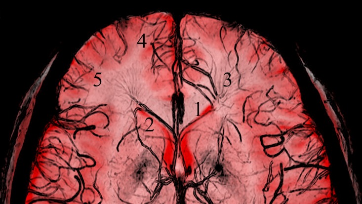
Fig 4. The fused image formed from T1-weighted and SW images.
The positional relationships between the anterior septal vein and the surrounding brain structures are clearly observed. (1, anterior septal vein; 2, anterior horn of lateral ventricle; 3, deep medullary veins; 4, superior frontal gyrus; 5, middle frontal gyrus).

