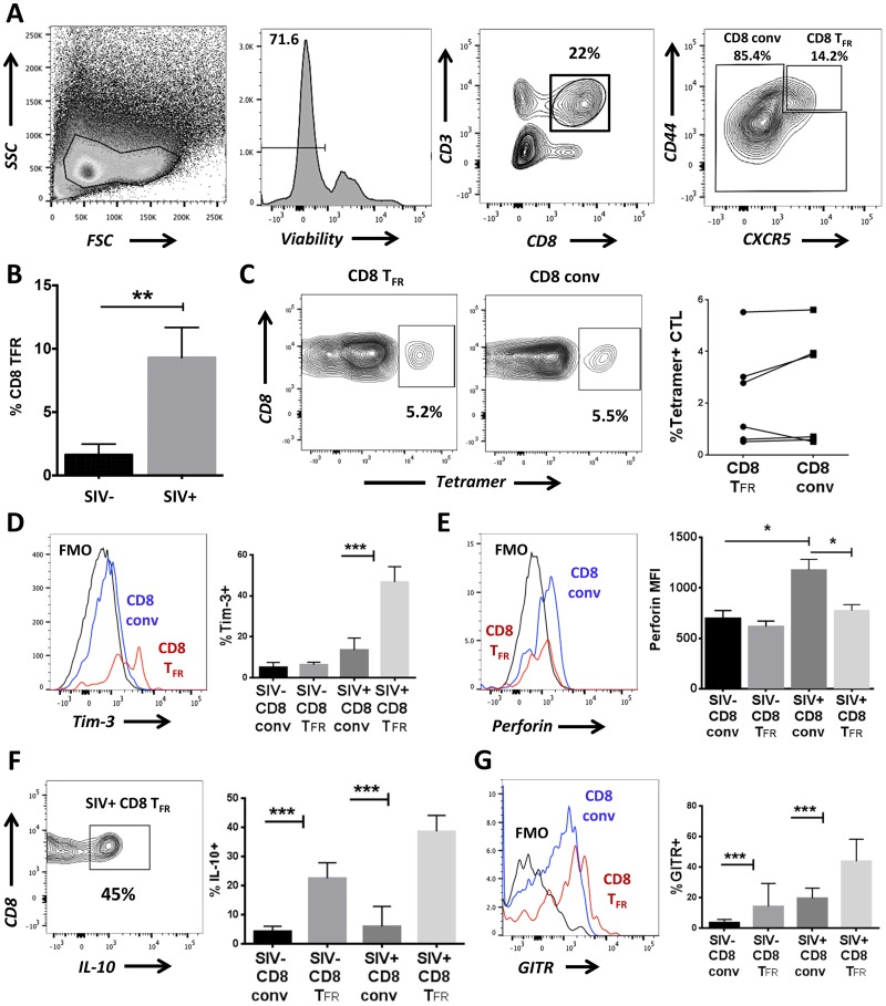
Fig 6. CD8 TFR are higher in SIV-infected than uninfected rhesus macaques.
Disaggregated cells from lymph nodes and spleen of SIV-uninfected (n = 6) and SIV-infected (n = 6) rhesus macaques were analyzed for CD8 TFR by flow cytometry. (A) Flow gating strategy to determine viable CD8 TFR (CD3+CD8+CXCR5hiCD44hi) and CD8 conv. (B) Percent CD8 TFR in SIV-uninfected compared to SIV-infected rhesus macaques. (C) Percent of SIV-Gag tetramer+ CD8 TFR compared to CD8 conv. CD8 TFR and CD8 conv from SIV-uninfected and SIV-infected rhesus macaques were analyzed for percent or MFI expression of (D) Tim-3, (E) perforin, (F) IL-10, and (G) GITR. Graphs depict median and range. Statistical significance was determined by non-parametric one-way ANOVA (Friedman test) and is displayed as * = p<0.05 and *** = p<0.001.

