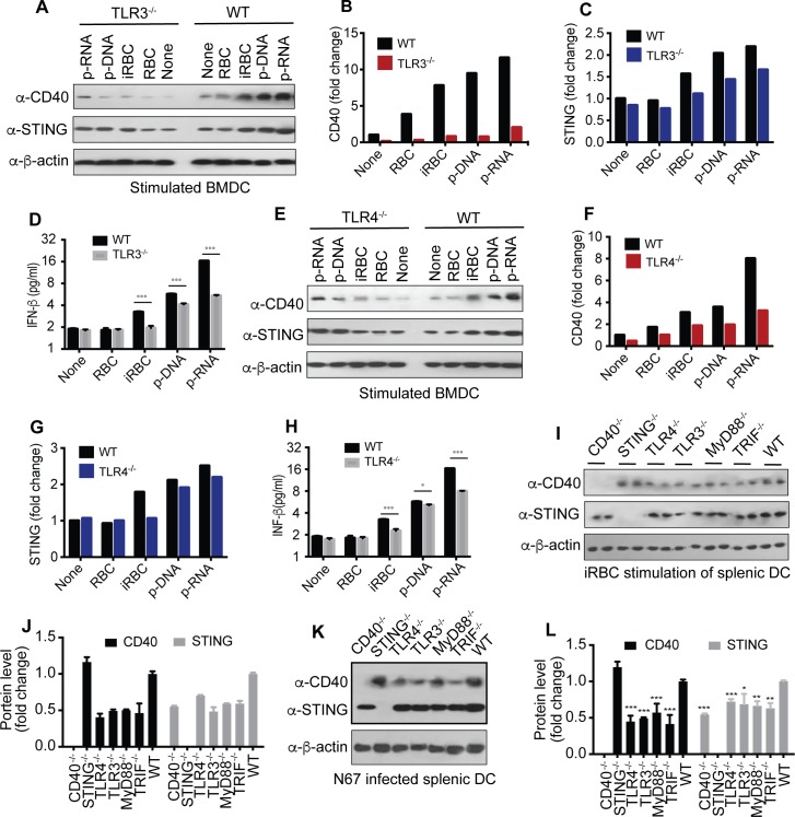
Fig 8. Effects of TLR signaling on CD40 and STING expressions in stimulated BMDCs and DCs from KO mice.
(A-D) CD40, STING, and IFN-β expressions in BMDCs from WT and TLR3-/- mice after stimulation. The cells were stimulated with the indicated agents for 24 h before harvesting proteins for Western blot. (A), Western blots; (B and C), quantitative signals of protein bands of CD40 and STING, respectively; (D) secreted IFN-β measured using ELISA. (E-H) the same as (A-D), except in TLR4-/- mice. (I and J) CD40 and STING expressions in purified splenic DCs from various KO mice after incubation with iRBCs for 24 h. Proteins from two mice each stimulated with iRBCs were separated on Western blot (I) and averaged signals were plotted in bar graphs (J). (K and L) CD40 and STING expressions in purified splenic DCs 4 days after infection with N67. Proteins were detected on Western blot (K) and scanned signals (means+s.d.) from three mice each were plotted (L). t-test, *P<0.05; **P<0.01; ***P<0.001. For all the experiments, 5 × 10⁵ cells were used.

