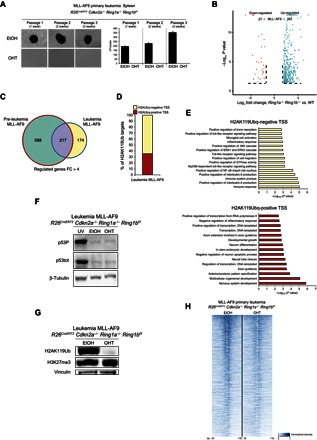
Fig. 5. Loss of H2AK119Ub in MLL-AF9–induced primary leukemia blocks cell growth.

(A) Methylcellulose colony assays starting from 1000 plated cells isolated from the spleen of a mouse that had developed a primary MLL-AF9 leukemia after intravenous inoculation of the MLL-AF9–transduced R26CreERT2 Cdkn2a−/− Ring1a−/− Ring1bf/f Lin− cells. Loss of PRC1 activity was induced by 4-OHT treatment at each methylcellulose passage. Quantifications of the colony number per plate are presented in the bottom panel. (B) Composite volcano plot showing the significantly differentially regulated genes 72 hours after 4-OHT treatment of the R26CreERT2 Cdkn2a−/− Ring1a−/− Ring1bf/f MLL-AF9 primary leukemia cells. EtOH was used as control treatment. (C) Overlaps between the up-regulated genes [fold change (FC) ≥ 4] in the MLL-AF9 preleukemic cells and MLL-AF9 leukemic cells upon loss of PRC1 activity. (D) Percentage of H2AK119Ub-enriched promoters undergoing transcriptional activation (fold change ≥ 4) after loss of PRC1 activity. Expression was determined by RNA-seq analysis in the indicated cells after 72 hours from the 4-OHT treatment. EtOH was used as control treatment. Red bars represent the percentage of H2AK119Ub-positive promoters (PRC1 direct targets); yellow bars represent the H2AK119Ub-negative promoters (PRC1 indirect targets). (E) P values of the significantly enriched pathways identified by gene ontology interrogation among the activated genes presented in (D). Top panel represents the functional pathways enriched among the PRC1 indirect targets; bottom panel represents the functional pathways enriched among the PRC1 direct targets. (F) Western blot analyses for total p53 (p53tot) and p53 phosphorylated (p53P) in the R26CreERT2 Cdkn2a−/− Ring1a−/− Ring1bf/f MLL-AF9 primary leukemia cells 72 hours after EtOH or 4-OHT addition, showing that the loss of PRC1 activity does not activate the p53 pathway. Ultraviolet-irradiated R26CreERT2 Cdkn2a−/− Ring1a−/− Ring1bf/f MLL-AF9 primary leukemia cells were used as p53/p53P-positive control (UV). β-Tubulin is presented as loading control. (G) Western blot analysis for H3K27me3 showing that its global deposition is not affected by the loss of H2AK119Ub in the R26CreERT2 Cdkn2a−/− Ring1a−/− Ring1bf/f MLL-AF9 primary leukemia cells 72 hours after EtOH or 4-OHT addition. Vinculin is presented as loading control. (H) Heat map representing the normalized H3K27me3 ChIP-seq intensities ±10 kb around the summit of H3K27me3-positive promoters identified in R26CreERT2 Cdkn2a−/− Ring1a−/− Ring1bf/f MLL-AF9 primary leukemia cells 72 hours after EtOH or 4-OHT addition.
