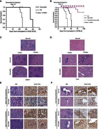
Fig. 6. PRC1 activity is essential for leukemia development both in vitro and in vivo.

(A) Kaplan-Meier survival curves of NOD/SCID mice intravenously inoculated with 1 × 10⁶ MLL-AF9 primary leukemic cells. Mice were treated with tamoxifen to inactivate the Ring1b allele at 12 days after leukemia transplant (arrow). For mice transplants: oil, n = 4; tamoxifen, n = 4. P value was determined by χ2 test. (B) Kaplan-Meier survival curves of C57BL/6 mice intravenously inoculated with 1 × 10⁶ MLL-AF9 primary leukemic cells. Mice were treated with tamoxifen to inactivate the Ring1b allele at 12 days after leukemia transplant (black arrowhead). For mice transplants: oil, n = 8; tamoxifen, n = 9. The pink arrowheads indicate the weekly tamoxifen injections for the group of mice (n = 5) that show improvement in the survival rate (pink survival curve), demonstrating that the leukemic cells that kill the mice are PRC1-proficient escapee cells. P value was determined by χ2 test. (C and D) H&E staining of spleen (C) and liver tissues (D) collected at day 6 or 30 after injection shows a milder leukemic phenotype in the tamoxifen-treated mice. (E and F) Immunohistochemical analyses of spleen (E) and liver tissues (F) collected at days 6 and 30 after tamoxifen injections, showing the rapid impairment of the active H2AK119Ub deposition in infiltrated leukemic cells compared to the control tissues (6 days after injection; top panels) as a consequence of the acute inactivation of PRC1 activity. Thirty days after tamoxifen injections, the infiltrated leukemic cells display an H2AK119Ub-positive staining (bottom panels), indicating the counterselection of H2AK119Ub-negative cells, with respect to the infiltrated leukemic cells with unexcised Ring1b allele. The far right panels are a magnification of the dashed square areas.
