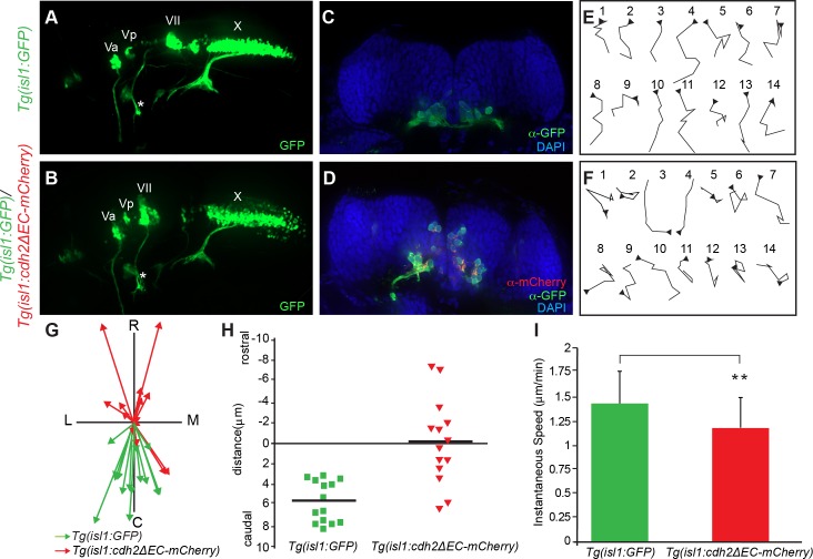
Fig 3. Inactivation of Cadherin-2 affects directionality of FBMN migration.
(A,B) Live imaging of lateral views of the hindbrain showing the positioning of CBMNs and their peripheral axonal projections at 48 hpf. (A) In wild-type Tg(isl1:GFP) embryos, FBMNs (VII) migration is complete and their axons (asterisk) project into the second branchial arch. (B) In Tg(isl1:GFP)/Tg(isl1:cdh2ΔEC-mCherry)vc25 embryos, the facial axons (asterisk) and trigeminal (Va,Vp), and vagus (X) axons can be seen to project normally. However, the FBMNs (VII) remain in r4 and lie in an abnormal dorsal position. (C,D) Coronal sections of 48hpf hindbrain from Tg(isl1:GFP) embryos at the level of r6 and Tg(isl1:GFP)/Tg(isl1:cdh2ΔEC-mCherry)vc25 embryos at the level of r4. FBMNs (α-GFP, green) in control Tg(isl1:GFP) embryos occupy a ventral position within r6. Cdh2ΔEC-mCherry-expressing FBMNs (α-mCherry, red) are found ectopically in a dorsal portion within r4. Nuclei are labeled with DAPI (blue). (E,F) Tracings of migratory paths of FBMNs captured from time-lapse images between 20–24 hpf from Tg(isl1:GFP) and Tg(isl1:GFP)/Tg(isl1:cdh2ΔEC-mCherry)vc25 embryos. Each time-lapse lasted 35 minutes with one frame every 5 minutes. Each trace is oriented so that caudal is to the bottom and medial is to the right. Arrowheads indicate the starting point for each cell. (G) Plot of the migratory tracks from start to endpoint shows a highly directional caudal migration of wild-type FBMNs (green arrows) in comparison to the random paths taken by Cdh2ΔEC-mCherry-expressing FBMNs (red arrows). C, caudal; R, rostral; L, lateral; M, medial (H) Quantitation of average distance traveled along the rostral-caudal axis by FBMNs in Tg(isl1:GFP) and Tg(isl1:GFP)/Tg(isl1:cdh2ΔEC-mCherry)vc25 embryos during the time-lapse sequences. (I) Quantitation of average instantaneous speed of FBMN movements in Tg(isl1:GFP) and Tg(isl1:GFP)/Tg(isl1:cdh2ΔEC-mCherry)vc25 embryos. (Mean values ± SD are shown; p < 0.05).

