
Figure 2. Effects of Wnt antagonists on Pk3 polarization in the epidermal ectoderm.
Embryos were injected with RNAs encoding GFP-Pk3 (150 pg), Xenopus HA-Vangl2 (120 pg), and LacZ (1 ng, A) or DN-Wnt11 (2 ng, B) or Ror2-TM (1 ng, C). (A–C) GFP fluorescence is shown in the epidermal ectoderm of embryos fixed at stage 15. Anterior is to the top. Scale bar, 20 µm. (D) Protein levels of GFP-Pk3 and HA-Vangl2 in the ectoderm analyzed by immunoblotting. A non-specific band detected by anti-HA antibody reflects protein loading.

Figure 2—figure supplement 1. ECD8 disrupts Pk3 polarization in the epidermis.
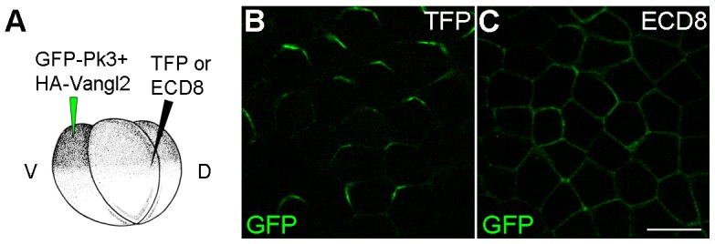
Four-cell embryos were injected at the ventral animal location with RNAs encoding GFP-Pk3 (150 pg) and Xenopus HA-Vangl2 (60 pg), followed by injection of RNAs for TurboFP635 (TFP, 1 ng) (B) or the extracellular domain of Frizzled 8 (ECD8, 1 ng) (C) into the dorsal marginal zone. GFP fluorescence in the epidermal ectoderm of stage 15 embryos is shown. Scale bar, 20 µm.