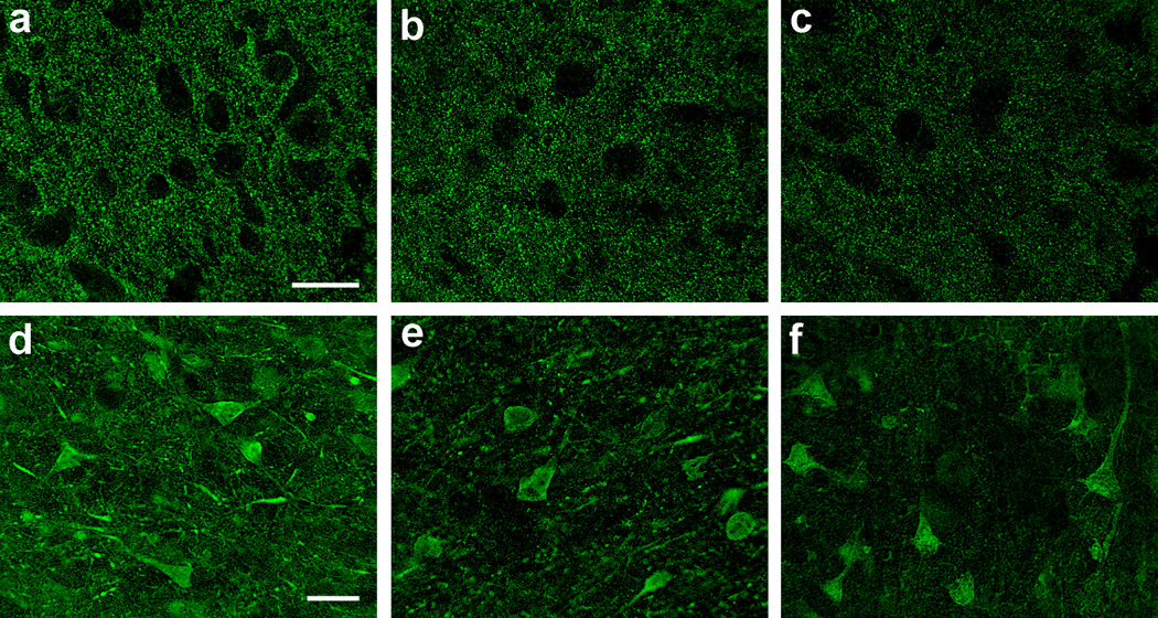
Fig. 4.
Immunofluorescence confocal microscopy for synaptophysin (SYP, a–c) and microtubule-associated protein-2 (MAP2, d–f) in the frontal cortical gray matter of HIV-infected adults. Shown are mild (a), moderate (b), and marked (c) loss of SYP immunoreactivity in neuropil between neuronal outlines, as are mild (d), moderate (e), and marked (f) loss of MAP2 immunoreactivity in neuronal soma and dendrites; scale bars: 20 µm (for SYP and MAP2)

