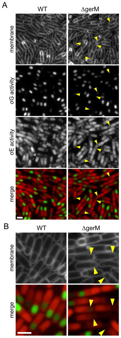
Figure 1. GerM is required for σG activity and forespore morphology.
A. Representative images of wild-type (WT, BCR1071) and ΔgerM (BAM833) sporulating cells at hour 3.5 (T3.5) of sporulation. Images (from top to bottom) are membrane staining with TMA-DPH, σG activity (PsspB-cfp), σE activity (PspoIID-mCherry) and merge of σG activity (green) and σE activity (red). Small and/or collapsed forespores with reduced σG activity are highlighted (yellow carets). Scale bar indicates 2 μm. A complete sporulation time-course comparing wild-type and ΔgerM can be found in Figure S1. B. Larger images highlighting the defects in σG activity and forespore morphology in the ΔgerM mutant.

