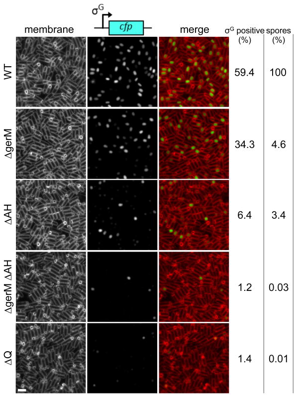
Figure 2. Synergistic defects in the gerM AH double mutant.
Representative images of sporulating cells harboring the σG-dependent reporter PsspB-cfp at hour 4 after the onset of sporulation. Images are wild-type (WT, BTD1609), ΔgerM (BCR1190), ΔAH (BCR1233), ΔgerM ΔAH double mutant (BCR1200), and ΔQ (BCR151). TMA-DPH-stained membranes (left), σG activity (middle) and a merged image (right) are shown. Scale bar represents 2 μm. The percentage of σG positive cells (n>600) at hour 4 are shown (see Materials and Methods for details). Spore titers relative to wild-type at hour 30 are indicated on the right. The data are representative of two biological replicates.

