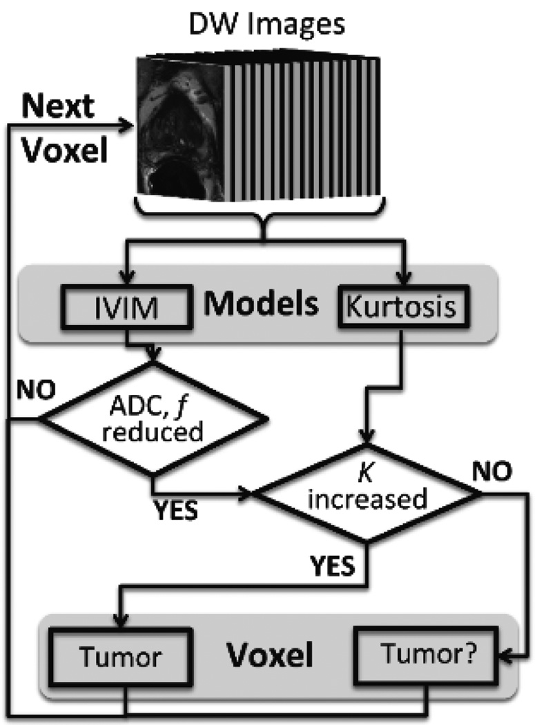
Figure 1.
Flowchart for the unsupervised multi-characteristic framework for prostate cancer localization. This technique utilizes DW-MRI images derived from a 16 b-value DW-MRI as an input to the diffusion models. In this work, two well-established models, intravoxel incoherent motion (IVIM) and kurtosis, were implemented. However, this framework has the capability of being extended to other MRI modalities and corresponding models. Based on previously reported findings for diffusion and kurtosis in prostate cancer tissue, certain criteria were used as shown in this figure to select tumor and tumor suspicious (“Tumor?”) voxels. This process was repeated on a voxel-by-voxel basis.

