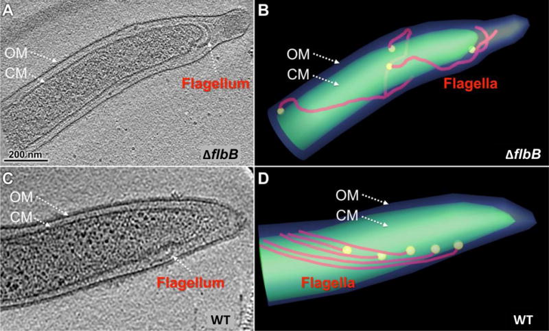
Figure 3. Periplasmic flagellar orientation in wild-type and ΔflbB mutant.

(A) A representative tomographic slice of a ΔflbB cell showing that the periplasmic flagella are abnormally oriented toward the cell pole. (B) A cartoon model of the ΔflbB mutant shown in (A) clearly illustrated the abnormal tilting of the flagella. (C) A representative tomographic slice of a WT cell showing the periplasmic flagella that are extended toward the cell body but not the cell pole. (D) A cartoon model of the WT cell showing the normal orientation of the flagella toward the cell body.
