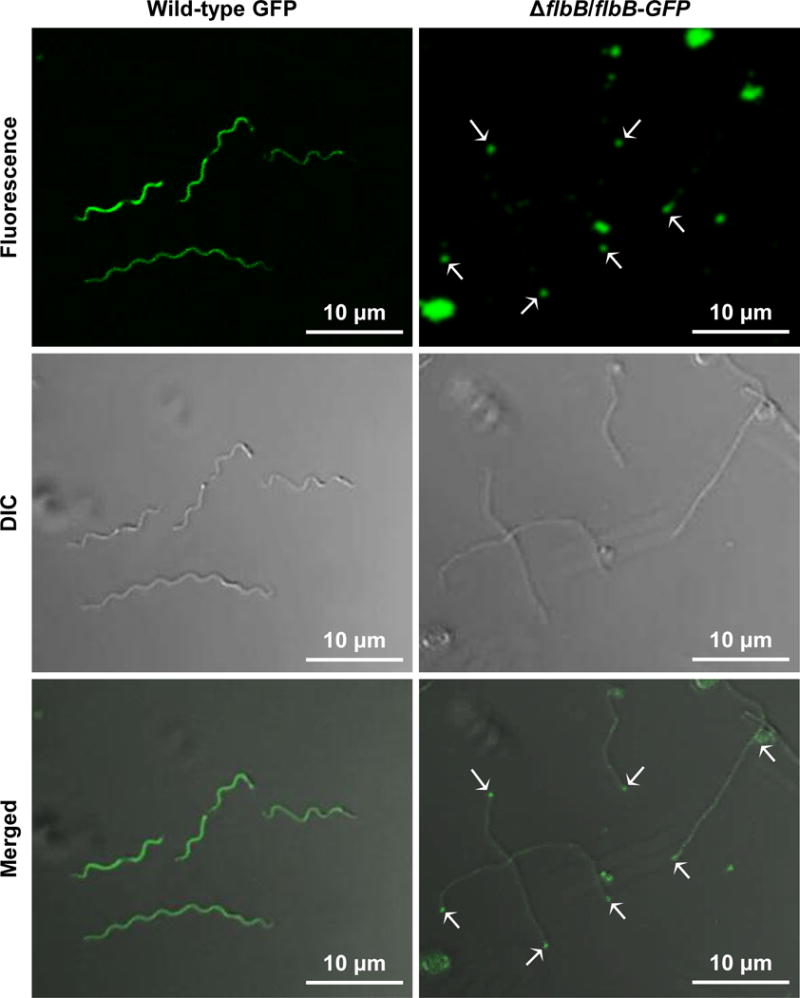
Figure 6. Expression and location of FlbB-GFP in B. burgdorferi.

Confocal microscopy showing the fluorescence (top), differential interference contrast (DIC; middle), and merged (bottom) micrographs of the wild-type cells expressing GFP (wild-type GFP) and of ΔflbB cells expressing FlbB-GFP (ΔflbB/flbB-GFP) at 64×. The white arrows indicate the location of FlbB-GFP in the ΔflbB/flbB-GFP cell tips (FlbB-GFP clusters were detected in approximately 73% cells tips). Even distribution of the GFP signal was observed throughout the wild-type GFP cells, as expected.
