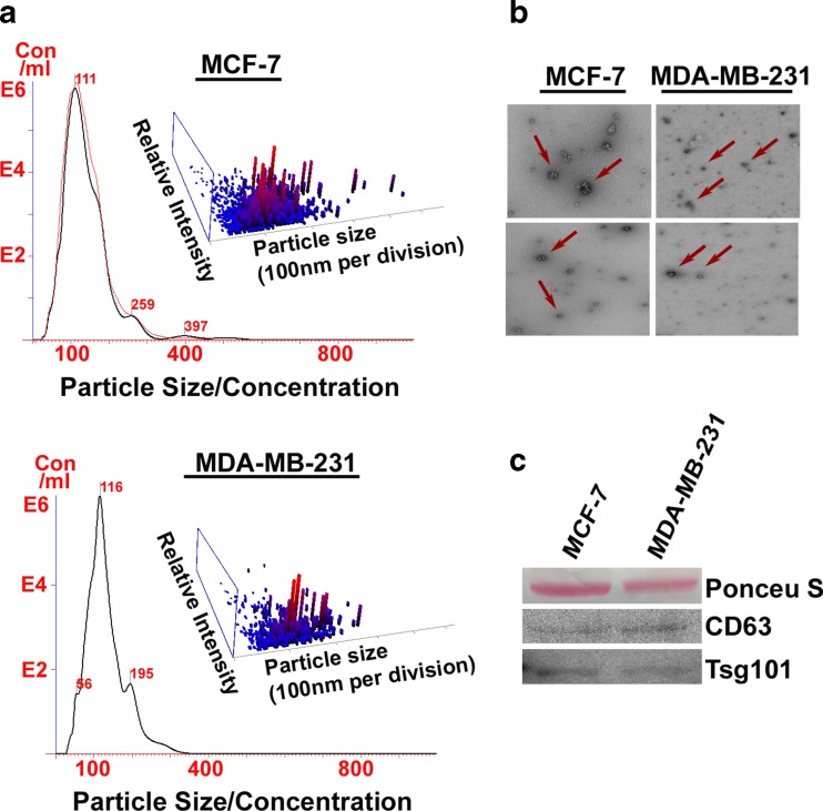
Fig. 2.
Qualitative and quantitative analysis of exosome in MCF-7 and MDA-MB-231 breast cancer cells using standard methods applied in exosome analysis. a Nano sight analysis showed different particle sizes with a peak in the desired range (exosomes). b Electron microscopy (TEM) confirmed the presence of exosomes based on morphology and size (Red arrows). c CD63 and TSG-101, markers for exosomes were expressed in the protein extract

