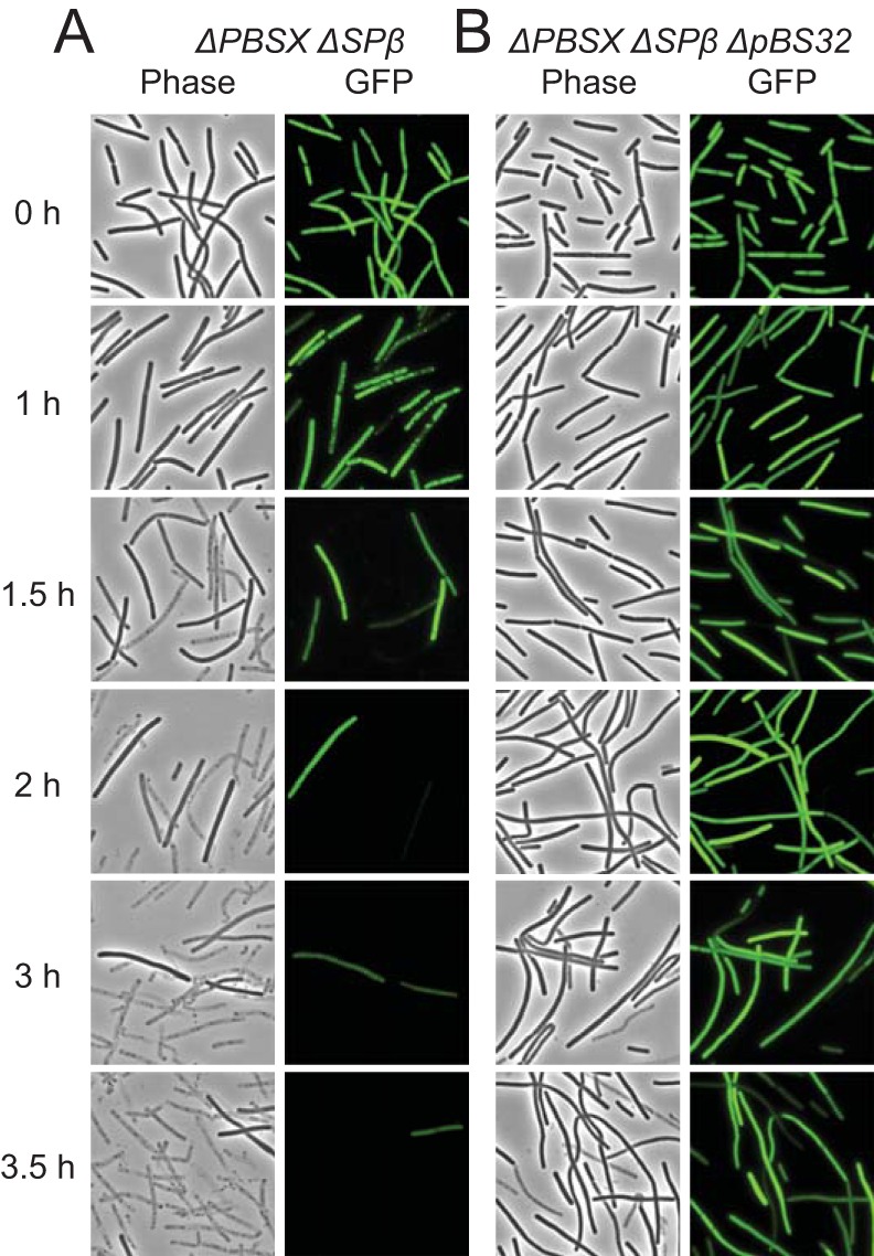
FIG 3.

pBS32 induction causes cell lysis. Phase-contrast and epifluorescence microscopy of GFP-expressing strains DK1233 (A) and DK1234 (B) at the indicated time points following treatment with 1 μg/ml mitomycin C at an OD600 of 0.1 (0 h). Strains were grown in the presence of 1 mM IPTG to induce GFP fluorescence. GFP fluorescence was false colored in green.
